. 2010 Jan;38(2):477-87.
doi: 10.1093/nar/gkp976. Epub 2009 Nov 11.

Cohesin promotes the repair of ionizing radiation-induced DNA double-strand breaks in replicated chromatin

Affiliations

Cohesin promotes the repair of ionizing radiation-induced DNA double-strand breaks in replicated chromatin

Christina Bauerschmidt et al. Nucleic Acids Res. 2010 Jan.

Abstract

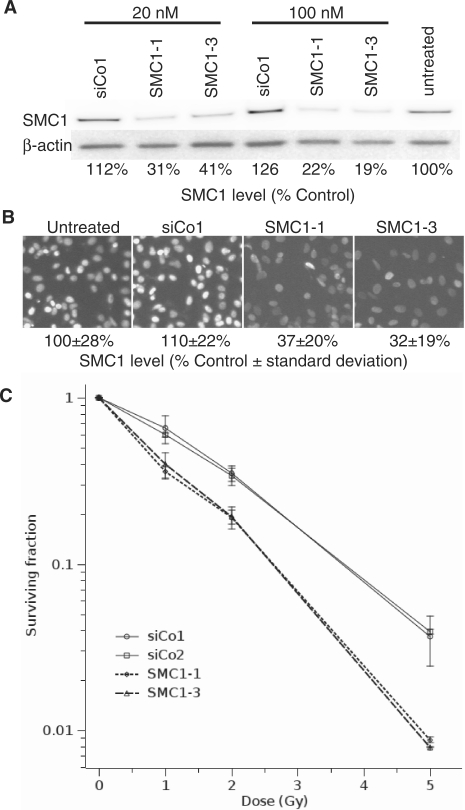
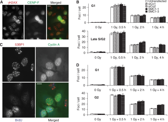
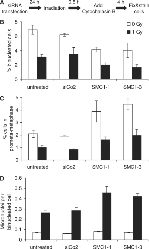
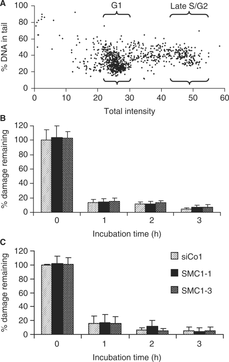
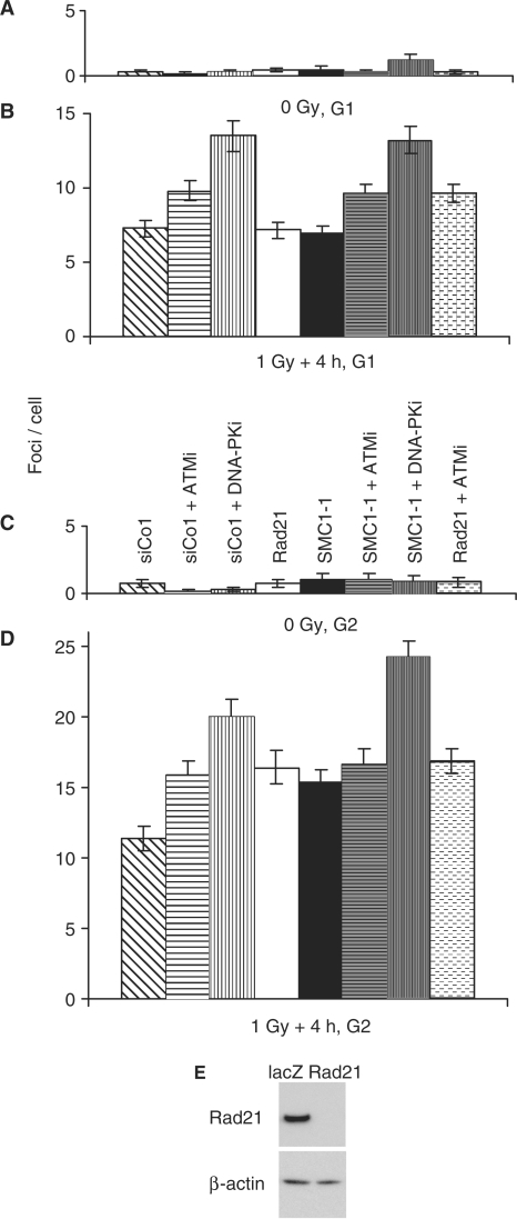
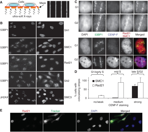
The cohesin protein complex holds sister chromatids together after synthesis until mitosis. It also contributes to post-replicative DNA repair in yeast and higher eukaryotes and accumulates at sites of laser-induced damage in human cells. Our goal was to determine whether the cohesin subunits SMC1 and Rad21 contribute to DNA double-strand break repair in X-irradiated human cells in the G2 phase of the cell cycle. RNA interference-mediated depletion of SMC1 sensitized HeLa cells to X-rays. Repair of radiation-induced DNA double-strand breaks, measured by gammaH2AX/53BP1 foci analysis, was slower in SMC1- or Rad21-depleted cells than in controls in G2 but not in G1. Inhibition of the DNA damage kinase DNA-PK, but not ATM, further inhibited foci loss in cohesin-depleted cells in G2. SMC1 depletion had no effect on DNA single-strand break repair in either G1 or late S/G2. Rad21 and SMC1 were recruited to sites of X-ray-induced DNA damage in G2-phase cells, but not in G1, and only when DNA damage was concentrated in subnuclear stripes, generated by partially shielded ultrasoft X-rays. Our results suggest that the cohesin complex contributes to cell survival by promoting the repair of radiation-induced DNA double-strand breaks in G2-phase cells in an ATM-dependent pathway.


Figures

Figure 1.
Figure 1.
SMC1 depletion radiosensitizes HeLa cells. (A) Depletion of SMC1 in HeLa cells using RNA interference. Cells were transfected with two different siRNAs against SMC1 (SMC1-1 and SMC1-3), a non-targeting siRNA (siCo1) or untransfected. Protein levels were analysed by western blotting. The percentage of SMC1 levels with respect to the untransfected control is shown. (B) Immunofluorescence microscopic analysis of SMC1 depletion. Details as in (A). (C) Colony formation assay for control and SMC1-depleted cells. Cells were transfected with two different siRNAs against SMC1 (SMC1-1 and SMC1-3) and two different non-targeting siRNAs (siCo1 and siCo2). Error bars are standard errors of the mean from three experiments.
Figure 2.
Figure 2.
SMC1 depletion impairs the repair of DSBs in G2-phase cells. (A) γH2AX and CENP-F staining in SMC1-depleted cells 2 h after 1 Gy X-irradiation. (B) Time course for the loss of 1 Gy X-ray-induced γH2AX foci in G1- (CENP-F negative) and late S/G2-phase cells (CENP-F strongly positive). (C) 53BP1, Cyclin A and BrdU staining in SMC1-depleted cells 2 h after 1 Gy X-irradiation. (D) Time course for the loss of 1 Gy X-ray-induced 53BP1 foci in G1 (BrdU-negative cyclin A-negative) and G2-phase cells (BrdU-negative cyclin A positive). Error bars are standard errors of the mean from two to three experiments.
Figure 3.
Figure 3.
SMC1 depletion affects progression through mitosis and enhances micronuclei induction. (A) Schematic representation of the experimental protocol. (B) Percentage of binucleated cells, (C) mitotic index and (D) numbers of micronuclei per binucleated cell obtained for different siRNA treatments following 0 or 1 Gy X-irradiation. Error bars are standard deviations from two experiments. The following numbers of cells were analysed in total for each data point (from left to right): 3366, 4290, 3654, 4566, 4877, 5211, 4718 and 5926.
Figure 4.
Figure 4.
Comet analysis of DNA strand break induction and repair. (A) Percentage of DNA in tail versus total DNA signal in a 4 Gy X-irradiated sample. The areas gated for the analysis of G1 and late S/G2-phase cells are indicated. (B and C) Time course for the percentage of damage remaining in G1 and late S/G2-phase cells following 4 Gy X-irradiation. Error bars are standard deviations from two experiments.
Figure 5.
Figure 5.
Contribution of DNA damage kinases and/or the cohesin factors SMC1 and Rad21 to 53BP1 foci loss in G1 and late S/G2-phase cells. 1 Gy X-ray-induced 53BP1 foci were scored in G1- (CENP-F negative) and late S/G2-phase cells (CENP-F strongly positive) 4 h after 1 Gy X-irradiation. (A and B) Un-irradiated and irradiated G1-phase cells. (C and D) Un-irradiated and irradiated late S/G2-phase cells. Error bars are standard deviations from two experiments. (E) Depletion of Rad21 using RNA interference. Cells were transfected with siRNAs against Rad21 or non-targeting siRNA (lacZ). Protein levels were analysed by western blotting. Rad21-depleted cells contained less than 2% of the levels found in control cells.
Figure 6.
Figure 6.
Recruitment of cohesin to ionizing radiation-induced DNA double strand breaks. (A) Schematic representation of micro-irradiation setup (left) and bright field image of the grid with 1 μm wide gaps/9 μm wide shielding used for generating defined patterns of DNA damage within the nucleus (right). (B) HeLa cells were grown on Mylar foil and irradiated with soft X-rays. After 1 h incubation, cells were pre-extracted with 0.1% Triton X-100, fixed and stained with the indicated antibodies. Each image is 125 μm wide. (C) As in (B) but cells were furthermore stained with CENP-F to determine the cell cycle phase. Each image is 15 μm wide. (D) Quantitative analysis of Rad21 and SMC1 stripe formation. Cells were divided into three groups of no or weak, medium and strong CENP-F staining. Microbeam tracks were predicted using the Rad21 or SMC1 signal, respectively, and then tested for co-localization with the 53BP1 signal. (E) Immunofluorescence microscopic analysis of Rad21 depletion. Control siRNA-transfected cells labelled with CellTrackerTM Green CMFDA (Invitrogen) were mixed with Rad21 siRNA-treated cells and stained for Rad21.

References

    1. Jackson SP. Sensing and repairing DNA double-strand breaks. Carcinogenesis. 2002;23:687–696. - PubMed
    1. Rothkamm K. Different means to an end: DNA double-strand break repair. In: Kiefer J, editor. Life Science and Radiation: Accomplishments and Future Directions. Berlin Heidelberg: Springer; 2004. pp. 179–186.
    1. Rothkamm K, Krüger I, Thompson LH, Löbrich M. Pathways of DNA double-strand break repair during the mammalian cell cycle. Mol. Cell. Biol. 2003;23:5706–5715. - PMC - PubMed
    1. Hinz JM, Yamada NA, Salazar EP, Tebbs RS, Thompson LH. Influence of double-strand-break repair pathways on radiosensitivity throughout the cell cycle in CHO cells. DNA Repair. 2005;4:782–792. - PubMed
    1. Saleh-Gohari N, Helleday T. Conservative homologous recombination preferentially repairs DNA double-strand breaks in the S phase of the cell cycle in human cells. Nucleic Acids Res. 2004;32:3683–3688. - PMC - PubMed

Publication types

MeSH terms