

Two distinct regions in Staphylococcus aureus GatCAB guarantee accurate tRNA recognition
- PMID: 19906721
- PMCID: PMC2811023
- DOI: 10.1093/nar/gkp955
Two distinct regions in Staphylococcus aureus GatCAB guarantee accurate tRNA recognition
Abstract
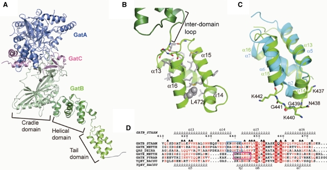
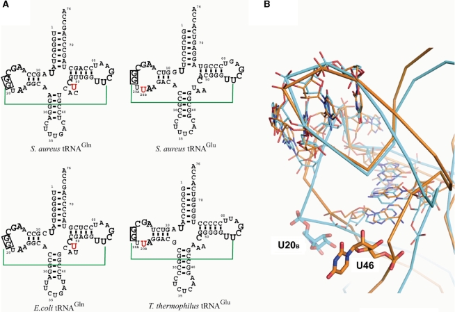
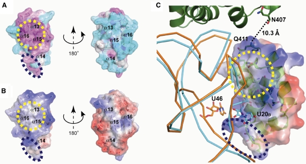
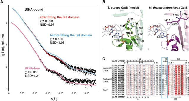
In many prokaryotes the biosynthesis of the amide aminoacyl-tRNAs, Gln-tRNA(Gln) and Asn-tRNA(Asn), proceeds by an indirect route in which mischarged Glu-tRNA(Gln) or Asp-tRNA(Asn) is amidated to the correct aminoacyl-tRNA catalyzed by a tRNA-dependent amidotransferase (AdT). Two types of AdTs exist: bacteria, archaea and organelles possess heterotrimeric GatCAB, while heterodimeric GatDE occurs exclusively in archaea. Bacterial GatCAB and GatDE recognize the first base pair of the acceptor stem and the D-loop of their tRNA substrates, while archaeal GatCAB recognizes the tertiary core of the tRNA, but not the first base pair. Here, we present the crystal structure of the full-length Staphylococcus aureus GatCAB. Its GatB tail domain possesses a conserved Lys rich motif that is situated close to the variable loop in a GatCAB:tRNA(Gln) docking model. This motif is also conserved in the tail domain of archaeal GatCAB, suggesting this basic region may recognize the tRNA variable loop to discriminate Asp-tRNA(Asn) from Asp-tRNA(Asp) in archaea. Furthermore, we identified a 3(10) turn in GatB that permits the bacterial GatCAB to distinguish a U1-A72 base pair from a G1-C72 pair; the absence of this element in archaeal GatCAB enables the latter enzyme to recognize aminoacyl-tRNAs with G1-C72 base pairs.
Figures
Similar articles
-
The Helicobacter pylori amidotransferase GatCAB is equally efficient in glutamine-dependent transamidation of Asp-tRNAAsn and Glu-tRNAGln.J Biol Chem. 2007 Apr 20;282(16):11866-73. doi: 10.1074/jbc.M700398200. Epub 2007 Feb 28. J Biol Chem. 2007. PMID: 17329242
-
Co-evolution of the archaeal tRNA-dependent amidotransferase GatCAB with tRNA(Asn).FEBS Lett. 2007 Jan 23;581(2):309-14. doi: 10.1016/j.febslet.2006.12.033. Epub 2007 Jan 2. FEBS Lett. 2007. PMID: 17214986 Free PMC article.
-
Methanothermobacter thermautotrophicus tRNA Gln confines the amidotransferase GatCAB to asparaginyl-tRNA Asn formation.J Mol Biol. 2008 Mar 28;377(3):845-53. doi: 10.1016/j.jmb.2008.01.064. Epub 2008 Jan 31. J Mol Biol. 2008. PMID: 18291416 Free PMC article.
-
Indirect tRNA aminoacylation during accurate translation and phenotypic mistranslation.Curr Opin Chem Biol. 2017 Dec;41:114-122. doi: 10.1016/j.cbpa.2017.10.009. Epub 2017 Nov 15. Curr Opin Chem Biol. 2017. PMID: 29156229 Review.
-
tRNA-dependent amino acid transformations.Nucleic Acids Symp Ser. 1997;(36):2-4. Nucleic Acids Symp Ser. 1997. PMID: 9478189 Review.
Cited by
-
Forward Genetics Reveals a gatC-gatA Fusion Polypeptide Causes Mistranslation and Rifampicin Tolerance in Mycobacterium smegmatis.Front Microbiol. 2020 Sep 24;11:577756. doi: 10.3389/fmicb.2020.577756. eCollection 2020. Front Microbiol. 2020. PMID: 33072044 Free PMC article.
-
The archaeal transamidosome for RNA-dependent glutamine biosynthesis.Nucleic Acids Res. 2010 Sep;38(17):5774-83. doi: 10.1093/nar/gkq336. Epub 2010 May 10. Nucleic Acids Res. 2010. PMID: 20457752 Free PMC article.
-
Neutron crystallographic study of heterotrimeric glutamine amidotransferase CAB.Acta Crystallogr F Struct Biol Commun. 2019 Mar 1;75(Pt 3):193-196. doi: 10.1107/S2053230X19000220. Epub 2019 Feb 21. Acta Crystallogr F Struct Biol Commun. 2019. PMID: 30839294 Free PMC article.
-
Robust and accurate prediction of residue-residue interactions across protein interfaces using evolutionary information.Elife. 2014 May 1;3:e02030. doi: 10.7554/eLife.02030. Elife. 2014. PMID: 24842992 Free PMC article.
-
Two enzymes bound to one transfer RNA assume alternative conformations for consecutive reactions.Nature. 2010 Sep 30;467(7315):612-6. doi: 10.1038/nature09411. Nature. 2010. PMID: 20882017
References
Publication types
MeSH terms
Substances
Associated data
- Actions
