. 2010 Jan;38(3):e14.
doi: 10.1093/nar/gkp932. Epub 2009 Nov 11.

Specific local induction of DNA strand breaks by infrared multi-photon absorption

Affiliations

Specific local induction of DNA strand breaks by infrared multi-photon absorption

D Träutlein et al. Nucleic Acids Res. 2010 Jan.

Abstract

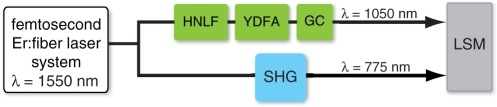
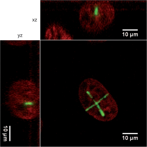
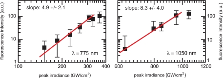
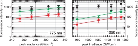
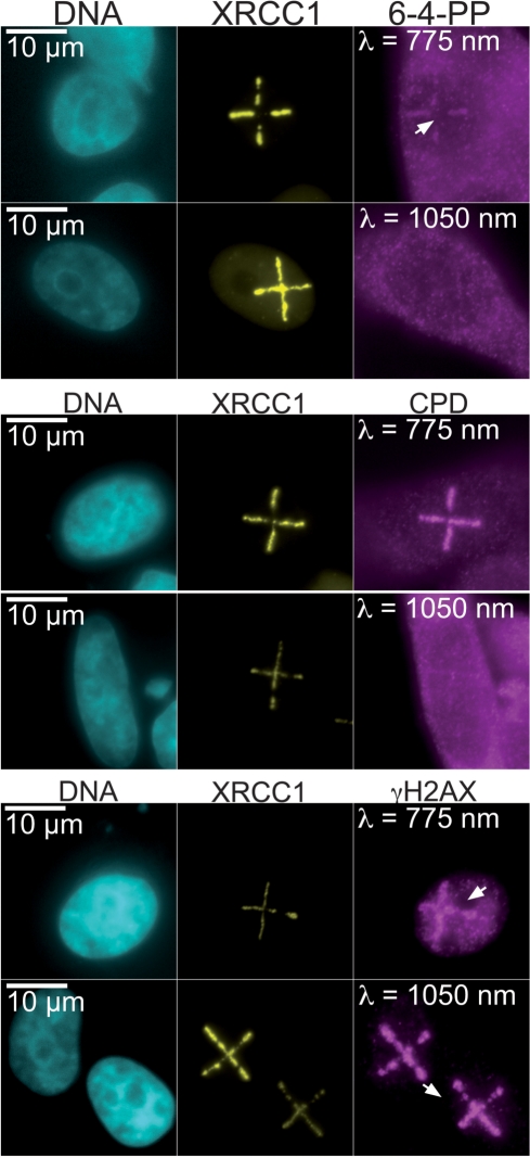
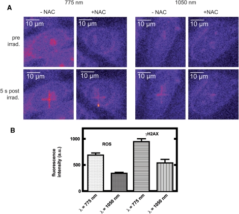
Highly confined DNA damage by femtosecond laser irradiation currently arises as a powerful tool to understand DNA repair in live cells as a function of space and time. However, the specificity with respect to damage type is limited. Here, we present an irradiation procedure based on a widely tunable Er/Yb : fiber femtosecond laser source that favors the formation of DNA strand breaks over that of UV photoproducts by more than one order of magnitude. We explain this selectivity with the different power dependence of the reactions generating strand breaks, mainly involving reactive radical intermediates, and the direct photochemical process leading to UV-photoproducts. Thus, localized multi-photon excitation with a wavelength longer than 1 microm allows for the selective production of DNA strand breaks at sub-micrometer spatial resolution in the absence of photosensitizers.


Figures

Figure 1.
Figure 1.
Schematic of the setup used for DNA microdamage, consisting of a femtosecond Er:fiber laser system, a periodically poled lithium niobate crystal for second harmonic generation (SHG), a highly nonlinear germanosilicate fiber (HNLF) for generation of a tailored supercontinuum and a Yb:fiber amplifier (YDFA) with subsequent grating compressor (GC). The microscope is a laser scanning confocal LSM 5 Pascal from Carl Zeiss.
Figure 2.
Figure 2.
XRCC1–GFP accumulates at irradiated sites in HeLa cell nuclei. HeLa cells expressing an XRCC1–GFP fusion were irradiated with the Er/Yb : fiber laser tuned to 1050 nm along two intersecting lines. The cells were fixed within 1 min after irradiation and imaged subsequently by confocal microscopy. The orthogonal projections highlight the spatial confinement of the XRCC1–GFP signal. Green: XRCC1–GFP. Red: Hoechst 33342.
Figure 3.
Figure 3.
Dependence of XRCC1 accumulation at 775 and 1050 nm from irradiance. Quantitative analysis of images of XRCC1–GFP expressing HeLa cells exposed to femtosecond pulses at 775 and 1050 nm as shown in Figure 2. Images were taken 64 s after irradiation at different peak irradiance values. The results are plotted on a double logarithmic scale. A linear function was fitted and the resulting slopes are 4.9 ± 2.1 at 775 nm and 8.3 ± 4.0 at 1050 nm. For each data point the accumulation of at least 10 cells was averaged. Error bars show standard deviation.
Figure 4.
Figure 4.
Dependence of signals specific for γH2AX (black circles), CPDs (green triangles) and 6-4 PPs (red squares) on the peak irradiance. Note that both axes are plotted on a logarithmic scale. Error bars show the standard deviation. Each data point represents the average value from at least eight cells. The slopes for a linear fit are 3.7 ± 0.9 (CPDs) and 3.8 ± 0.5 (6-4 PPs) at 775 nm, and 8.9 ± 2.6 (CPDs) and 8.1 ± 1.2 (6-4 PPs) at 1050 nm.
Figure 5.
Figure 5.
Femtosecond laser pulses at 1050 nm selectively induce DSBs in HeLa cell nuclei. Immunocytochemical analysis of HeLa cells expressing XRCC1–GFP. Cells were irradiated at 775 nm (253 GW/cm2) or 1050 nm (947 GW/cm2), fixed and labeled with antibodies specific for γH2AX, CPDs and 6-4 PPs. The XRCC1–GFP signal served as a positive control and allowed to identify the irradiated region.
Figure 6.
Figure 6.
ROS contribute to DNA strand breaks induced by femtosecond irradiation. (A) HeLa cells were incubated with CM-H2DCFDA before excitation at 775 nm (326 GW/cm2) and 1050 nm (800 GW/cm2). Images taken prior to and immediately after exposure are shown. As a control, cells were pre-treated with 4 mM NAC. NAC effectively quenches the ROS-specific signal. (B) Irradiation conditions were chosen such as to generate ∼50% less ROS at 1050 nm as compared to 775 nm. Cells were irradiated in parallel experiments, monitored for the production of ROS and labeled with a γH2AX-specific antibody. The effect on the γH2AX-specific signal correlates quantitatively with the decrease in ROS. Data points are an average of at least 34 cells. Error bars show standard error of the mean.

References

    1. Cremer C, Cremer T, Fukuda M, Nakanishi K. Detection of laser–UV microirradiation-induced DNA photolesions by immunofluorescent staining. Hum. Genet. 1980;54:107–110. - PubMed
    1. Cremer C, Cremer T, Jabbur G. Laser–UV-microirradiation of Chinese hamster cells: the influence of the distribution of photolesions on unscheduled DNA synthesis. Photochem. Photobiol. 1981;33:925–928. - PubMed
    1. Mone MJ, Volker M, Nikaido O, Mullenders LH, van Zeeland AA, Verschure PJ, Manders EM, van Driel R. Local UV-induced DNA damage in cell nuclei results in local transcription inhibition. EMBO Rep. 2001;2:1013–1017. - PMC - PubMed
    1. Volker M, Mone MJ, Karmakar P, van Hoffen A, Schul W, Vermeulen W, Hoeijmakers JHJ, van Driel R, van Zeeland AA, Mullenders LHF. Sequential assembly of the nucleotide excision repair factors in vivo. Mol. Cell. 2001;8:213–224. - PubMed
    1. Okano S, Lan L, Caldecott KW, Mori T, Yasui A. Spatial and temporal cellular responses to single-strand breaks in human cells. Mol. Cell Biol. 2003;23:3974–3981. - PMC - PubMed

Publication types

Substances