Comparative Study
. 2010 Feb;77(2):288-97.
doi: 10.1124/mol.109.059733. Epub 2009 Nov 11.

Role of helix 8 of the thyrotropin-releasing hormone receptor in phosphorylation by G protein-coupled receptor kinase

Affiliations
Comparative Study

Role of helix 8 of the thyrotropin-releasing hormone receptor in phosphorylation by G protein-coupled receptor kinase

Austin U Gehret et al. Mol Pharmacol. 2010 Feb.

Abstract

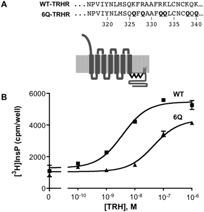
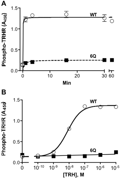
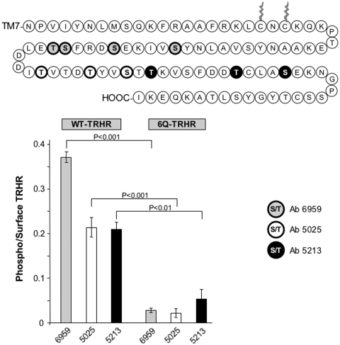
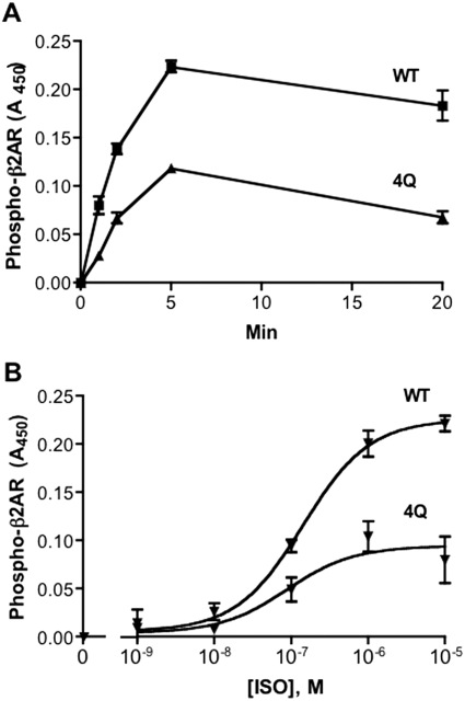
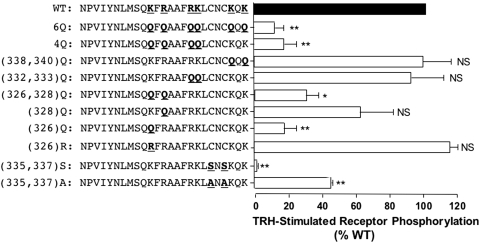
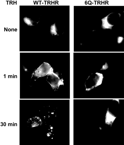
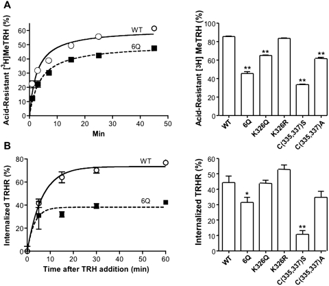
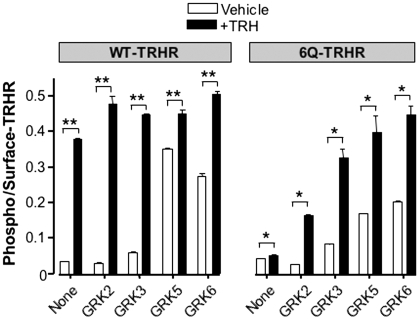
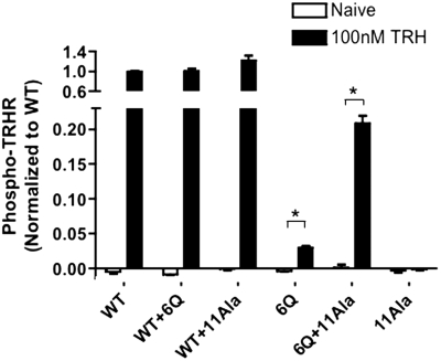
The thyrotropin-releasing hormone (TRH) receptor undergoes rapid and extensive agonist-dependent phosphorylation attributable to G protein-coupled receptor (GPCR) kinases (GRKs), particularly GRK2. Like many GPCRs, the TRH receptor is predicted to form an amphipathic helix, helix 8, between the NPXXY motif at the cytoplasmic end of the seventh transmembrane domain and palmitoylation sites at Cys335 and Cys337. Mutation of all six lysine and arginine residues between the NPXXY and residue 340 to glutamine (6Q receptor) did not prevent the receptor from stimulating inositol phosphate turnover but almost completely prevented receptor phosphorylation in response to TRH. Phosphorylation at all sites in the cytoplasmic tail was inhibited. The phosphorylation defect was not reversed by long incubation times or high TRH concentrations. As expected for a phosphorylation-defective receptor, the 6Q-TRH receptor did not recruit arrestin, undergo the typical arrestin-dependent increase in agonist affinity, or internalize well. Lys326, directly before phenylalanine in the common GPCR motif NPXXY(X)(5-6)F(R/K), was critical for phosphorylation. The 6Q-TRH receptor was not phosphorylated effectively in cells overexpressing GRK2 or in in vitro kinase assays containing purified GRK2. Phosphorylation of the 6Q receptor was partially restored by coexpression of a receptor with an intact helix 8 but without phosphorylation sites. Phosphorylation was inhibited but not completely prevented by alanine substitution for cysteine palmitoylation sites. Positively charged amino acids in the proximal tail of the beta2-adrenergic receptor were also important for GRK-dependent phosphorylation. The results indicate that positive residues in helix 8 of GPCRs are important for GRK-dependent phosphorylation.


Figures

Fig. 1.
Fig. 1.
Signaling by TRH receptors. A, sequence of the helix 8 region of the WT and mutant rat TRH receptors. Helix 8 is depicted in boldface type. B, HEK293 cells were transfected with DNA encoding WT or 6Q-TRH receptors with N-terminal V5 epitope tags. Cells were metabolically labeled overnight with [3H]inositol and incubated with 10 mM LiCl and vehicle or TRH for 30 min, when total [3H]inositol phosphates were quantified. TRH receptor density was measured by cell-surface ELISA and was identical for both receptors. Results are from a representative experiment performed in duplicate.
Fig. 2.
Fig. 2.
Rate and concentration-dependence of TRH-induced receptor phosphorylation. Cells were transfected with WT or 6Q-TRH receptors. A, cells were incubated with 100 nM TRH for the times shown. B, cells were incubated for 5 min with the concentrations of TRH shown. Cells were fixed, and phosphorylated receptor was measured by ELISA using antibody 6959. Receptor levels were 32% lower for 6Q than for WT receptors. Backgrounds were identical in mock-transfected cells and cells not exposed to TRH. Results show the mean and range of duplicates in a representative experiment.
Fig. 3.
Fig. 3.
Phosphorylation of different sites in the TRH receptor cytoplasmic tail. Cells were mock-transfected or were transfected with WT or 6Q-TRH receptor and then incubated for 5 min with vehicle or 100 nM TRH and fixed. Phosphorylation was measured with three different affinity-purified antibodies directed against phosphopeptides from the receptor tail (Jones et al., 2007). Antibodies were raised against peptides with the following residues phosphorylated: Ab6959: Ser355, Ser360, Ser364, and Thr365; Ab5025: Thr371, Thr375, and Ser378; and Ab5213: Thr380, Thr387, and Ser391. Results are normalized to correct for differences (<20%) in surface receptor expression and show mean and S.E.M. values of triplicates in a representative experiment. Signals in mock-transfected cells were equal to or slightly higher than signals in cells treated with vehicle, indicating that there was no constitutive phosphorylation.
Fig. 4.
Fig. 4.
Rate and concentration-dependence of isoproterenol-induced β2-adrenergic receptor phosphorylation. Cells were transfected with HA-tagged WT or 4Q-β2-adrenergic receptors. A, cells were incubated with 1 μM isoproterenol for the times indicated. B, cells were incubated for 5 min with the concentrations of isoproterenol (ISO) shown. Phosphorylated receptor was measured by ELISA using antibodies against phospho-β2-adrenergic receptor (pSer355/pSer356) and surface receptor expression with anti-HA antibody. 4Q receptors were expressed at 95% of the level of WT receptors. Results of two independent experiments performed in duplicate are shown.
Fig. 5.
Fig. 5.
Role of positively charged residues in helix 8 and palmitoylation sites. V5-tagged TRH receptors with the mutations shown were expressed in HEK293 cells, which were incubated with or without 100 nM TRH for 5 min. Phosphorylated and cell surface receptor levels were quantified by ELISA. For each receptor, the level of phosphorylation was normalized to the level of the cell surface receptor. Results are expressed relative to the amount of phosphorylated WT receptor in the same experiment. Shown are the mean ± S.E.M. values of results obtained in three or four experiments, each performed in duplicate or triplicate. ∗∗, P < 0.001, ∗, P < 0.01; NS, not significant versus WT.
Fig. 6.
Fig. 6.
Interaction of TRH receptors with arrestin3-GFP. HEK293 cells expressing arrestin3-GFP and either WT or 6Q-TRH receptor were stimulated with 1 μM TRH for the times shown, fixed, and imaged.
Fig. 7.
Fig. 7.
Internalization of TRH receptors and formation of an acid-resistant agonist-receptor complex. HEK293 cells were transfected with DNA encoding WT or mutant TRH receptors. A, cells were incubated with 5 nM [3H]MeTRH for the times shown (left) or with 10 nM [3H]MeTRH for 45 min (right), when the fraction of specifically bound hormone resistant to ice-cold 0.2 N acetic acid/0.5 M NaCl, pH 2.5, was measured. B, cells were stimulated with 100 nM TRH for the times shown (left) or with 1 μM TRH for 45 min (right), when the amount of receptor on the cell surface was measured by ELISA. Results represent the mean ± S.E.M. of four determinations. ∗, P < 0.05, ∗∗, P < 0.01 versus WT.
Fig. 8.
Fig. 8.
Effect of GRK overexpression. Cells were transfected with WT or 6Q TRH receptors together with empty vector or GRK2, 3, 5, or 6 and then stimulated with vehicle or 100 nM TRH for 5 min. Phosphorylated and cell-surface receptor were measured by ELISA, and phosphorylation values were normalized to receptor expression. Results show the mean and S.E.M. of triplicates in a representative experiment. ∗∗, P < 0.001, ∗, P < 0.01 versus vehicle-treated control.
Fig. 9.
Fig. 9.
In vitro phosphorylation of TRH receptors and interactions with GRK2. A, cells expressing WT or 6Q-TRH receptors with or without K220R-GRK2 were incubated with antibody to V5 to label surface receptors, washed, and incubated with or without 10 μM TRH for 0 to 5 min. Receptors were solubilized with dodecyl-β-d-maltoside, and immune complexes were collected on protein A/G beads. Immunoprecipitates were resolved on SDS-polyacrylamide gel electrophoresis and blotted for either total receptor (top) or GRK2 (bottom). B, cells were transfected with WT or 6Q receptors with or without GRK6 and exposed to vehicle or 100 nM TRH for 5 min. Proteins were solubilized with Triton X-100, and phosphorylated and total receptors were immunoprecipitated with antibodies to phosphoreceptor (Ab6959) (top) and V5 epitope (bottom), respectively, resolved on gels and blotted with anti-V5 antibody. C, membranes were isolated from cells expressing WT, 6Q, or K326Q-TRH receptors and incubated in in vitro kinase buffer with or without 100 nM TRH and 100 nM GRK2 for 5 min. Immunoprecipitation and immunoblotting were carried out as in B. Densitometric analysis of blots of total receptor in lysates from the same experiment (data not shown) revealed that 6Q and K326Q receptors were expressed at 92 and 65% of WT, respectively.
Fig. 10.
Fig. 10.
Complementation of TRH-stimulated receptor phosphorylation. HEK293 cells were cotransfected with DNA encoding two different TRH receptors, as shown; when only one receptor was expressed, a control plasmid was included. The 11Ala-TRH receptor was N-terminally tagged with HA, and all other receptors were tagged with V5 epitopes. Cells were incubated with or without 100 nM TRH for 5 min. Phosphorylated receptors were quantified by ELISA using antibody 6959. Signal from mock-transfected cells was subtracted from experimental values. The results are normalized to the phosphorylation of WT-TRH receptor in the same experiment. Coexpression of the 11Ala receptor increased surface expression of the WT and 6Q receptors by 21 and 31%, respectively. The increase in 6Q receptor expression does not account for the >500% increase in 6Q receptor phosphorylation caused by coexpression of 11Ala receptors. Shown are the mean ± S.E.M. values of three independent experiments performed in duplicate. ∗, P < 0.01.

Similar articles

Cited by

References

    1. Altenbach C, Kusnetzow AK, Ernst OP, Hofmann KP, Hubbell WL. (2008) High-resolution distance mapping in rhodopsin reveals the pattern of helix movement due to activation. Proc Natl Acad Sci U S A 105: 7439–7444 - PMC - PubMed
    1. Benovic JL, Onorato J, Lohse MJ, Dohlman HG, Staniszewski C, Caron MG, Lefkowitz RJ. (1990) Synthetic peptides of the hamster beta 2-adrenoceptor as substrates and inhibitors of the beta-adrenoceptor kinase. Br J Clin Pharmacol 30 (Suppl 1): 3S–12S - PMC - PubMed
    1. Cai K, Klein-Seetharaman J, Farrens D, Zhang C, Altenbach C, Hubbell WL, Khorana HG. (1999) Single-cysteine substitution mutants at amino acid positions 306–321 in rhodopsin, the sequence between the cytoplasmic end of helix VII and the palmitoylation sites: sulfhydryl reactivity and transducin activation reveal a tertiary structure. Biochemistry 38: 7925–7930 - PubMed
    1. Cole C, Barber JD, Barton GJ. (2008) The Jpred 3 secondary structure prediction server. Nucleic Acids Res 36: W197–W201 - PMC - PubMed
    1. Cook LB, Zhu CC, Hinkle PM. (2003) Thyrotropin-releasing hormone receptor processing: role of ubiquitination and proteasomal degradation. Mol Endocrinol 17: 1777–1791 - PubMed

Publication types

MeSH terms

Substances

LinkOut - more resources