. 2009 Dec;136(24):4165-76.
doi: 10.1242/dev.044263. Epub 2009 Nov 11.

Drosophila laminins act as key regulators of basement membrane assembly and morphogenesis

Affiliations

Drosophila laminins act as key regulators of basement membrane assembly and morphogenesis

Jose M Urbano et al. Development. 2009 Dec.

Abstract

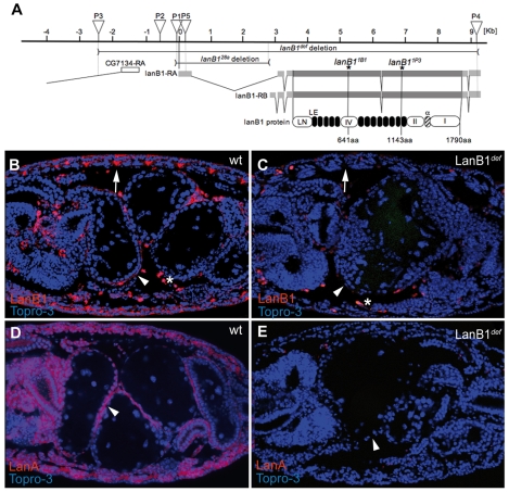
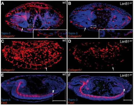
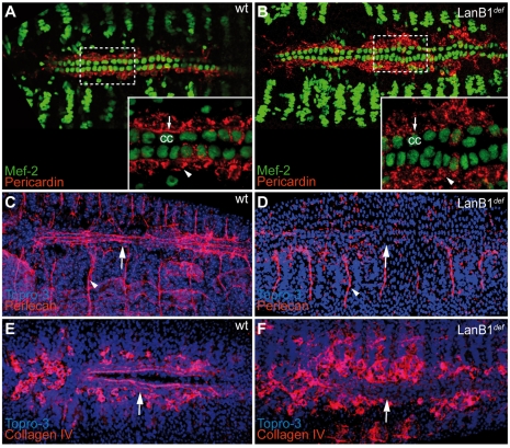
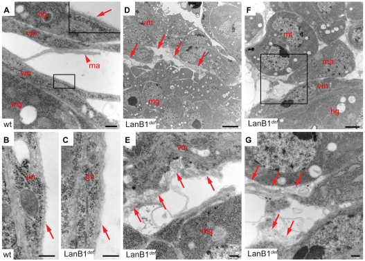
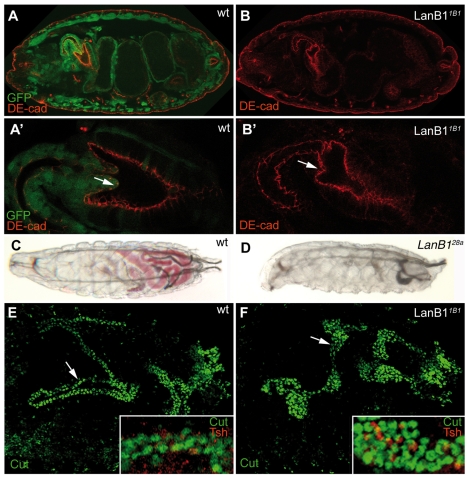
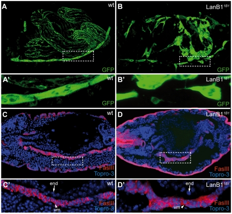
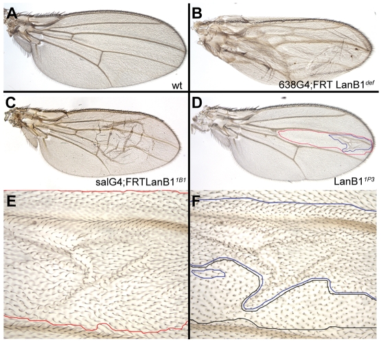
Laminins are heterotrimeric molecules found in all basement membranes. In mammals, they have been involved in diverse developmental processes, from gastrulation to tissue maintenance. The Drosophila genome encodes two laminin alpha chains, one beta and one Gamma, which form two distinct laminin trimers. So far, only mutations affecting one or other trimer have been analysed. In order to study embryonic development in the complete absence of laminins, we mutated the gene encoding the sole laminin beta chain in Drosophila, LanB1, so that no trimers can be made. We show that LanB1 mutant embryos develop until the end of embryogenesis. Electron microscopy analysis of mutant embryos reveals that the basement membranes are absent and the remaining extracellular material appears disorganised and diffuse. Accordingly, abnormal accumulation of major basement membrane components, such as Collagen IV and Perlecan, is observed in mutant tissues. In addition, we show that elimination of LanB1 prevents the normal morphogenesis of most organs and tissues, including the gut, trachea, muscles and nervous system. In spite of the above structural roles for laminins, our results unravel novel functions in cell adhesion, migration and rearrangement. We propose that while an early function of laminins in gastrulation is not conserved in Drosophila and mammals, their function in basement membrane assembly and organogenesis seems to be maintained throughout evolution.


Figures

Fig. 1.
Fig. 1.
Molecular description of LanB1 mutant alleles and distribution of the LanB1 protein. (A) Schematic representation of the LanB1 genomic region. Both laminin β transcripts (LanB1-RA and LanB1-RB) with their coding and untranslated regions are represented in dark and light grey horizontal bars, respectively. The white box represents the 5′ UTR of the gene CG7134. The localisations of transposon insertions appear as inverted triangles [P1=P(EP)EP2178, P2=EP600, P3=P{XP}d04880, P4=PBac{RB}LanB1e02263, P5=l(2)k05404]. The horizontal lines correspond to the genomic region deleted in the LanB128a allele and the deficiency LanB1def. Asterisks indicate the positions of stop codons in the LanB11B1 and LanB11P3 alleles. Conserved domains are schematised as follows: white, from left to right, LanB1 N-terminal domain (LN), domain IV, domain II and domain I; black, 13 laminin-type EGF-like domains (LE); and stripped, the alpha domain. In all figures, embryos are oriented with anterior to the left. (B) In stage 16 wild-type embryos, LanB1 (red) is found in BMs surrounding most tissues (arrowhead) and at muscle attachment sites (arrow). (C) This expression is lost in maternal and zygotic homozygous LanB1def mutant embryos. (D) In wild-type embryos, LanA (red) is found at BMs. (E) This expression is not detected in LanB1def mutant embryos. In all figures, the nuclear marker TO-PRO 3 is in blue. wt, wild type.
Fig. 2.
Fig. 2.
Localisation of LanB1 during imaginal and pupal wing development. (A-F‴) Vertical lines indicate the position of transverse sections shown in the accompanying panels. (A-A″) Basal side of a third instar wing disc stained with LanB1 (green) and Armadillo (red). LanB1 is localised at the basal side of the columnar epithelium and peripodial membrane. (B-B″) In 0- to 4-hour-APF pupal wings, LanB1 (green) and βPS integrin (red) colocalise at the basal surface of all wing epithelial cells. (C-D‴) This localisation persists from 20 to 28 hours APF. LanB1 also localises in the lacunae formed between the dorsal and ventral wing spaces from 20 to 44 hours (C-F‴). (E-F‴) However, from 32 to 44 hours APF, LanB1 and βPS integrin are found in mutually exclusive patterns, with LanB1 being found at the basal surface of veins (D″,F″) and βPS integrin at that of interveins (E″,F‴).
Fig. 3.
Fig. 3.
LanB1 is required for proper localisation of Collagen IV at basement membranes. (A-F) Dorsal views of stage 16 embryos (A,B). Lateral views of stage 15 (C,D) and 16 (E,F) embryos. (A,D) In wild-type embryos Collagen IV (red) is found at BMs surrounding the brain (arrow) and midgut (magnification in the white box). (B) This localisation is severely disrupted in LanB1def embryos. (C) In addition, Collagen IV localisation in the ventral nerve cord channels of a stage 15 embryo (arrow) is lost in LanB1def embryos (D, arrow). (E,F) In stage 16 embryos, the ventral nerve cord, visualised with anti-FasII antibody (red), fails to condense properly in LanB1def mutants. White horizontal bars indicate the length of CNS condensation.
Fig. 4.
Fig. 4.
LanB1 is essential for correct localisation of ECM components during heart morphogenesis. (A,B) Dorsal view of stage 16 embryos labelled with anti-Pericardin (red) and anti-Mef2 (green) antibodies. (A) In wild-type embryos, Pericardin is localised at the basal surface of cc (arrow) and around pericardial cells (arrowhead). This localisation is disrupted in LanB1def embryos (B). (C-F) Localisation of other ECM components, such as Perlecan (red in C and D) and Collagen IV (red in E and F), around the heart (arrows) is also altered in LanB1def embryos. Perlecan localisation at muscle attachments remains unaffected (C,D, arrowheads).
Fig. 5.
Fig. 5.
Ultrastructural analysis of embryos lacking LanB1. (A-G) Wild-type (A,B) and LanB1def mutant (C-G) embryos at early stage 17. (A) Basal surfaces of the midgut (mg) and hindgut that are closely associated with a layer of visceral musculature (vm). All tissue surfaces exposed to the haemolymph show a uniform layer of BM (arrow in inset). A lamellipodium of a macrophage is also seen that is not accumulating a BM (arrowhead). (B) The CNS is surrounded by a layer of perineural cells that is covered by BM in wild type. (C) In LanB1 mutant embryos only residual ECM material is detected. (D) Gaps between mg and vm cells are apparent in LanB1 mutants (arrows), often filled with unorganised and diffuse ECM material (E, arrows). (F) Section of Malphigian tubule (mt), hindgut with associated vm and a macrophage in LanB1 mutant embryo (F). (G) Close up of boxed region in F. Arrows point to diffuse ECM material at the surface of the mt and vm. Scale bars: 1 μm in F; 2 μm in D; 100 nm in A,C,E,G. hg, hindgut; ma, macrophage; mg, midgut; mt, Malphigian tubule; pn, perineural cell; vm, visceral musculature.
Fig. 6.
Fig. 6.
Elimination of LanB1 function affects the migration of different cell populations. (A-D) Embryos were labelled with anti-Hindsight (Hnt, brown) and anti-β-gal (blue) antibodies to visualise the midgut (mg) cells and to mark the balancer chromosome, respectively. (A,B) The mg primordia has completed its migration and met in the centre of stage 13 wild-type embryos (A, arrow). By contrast, migration is delayed in LanB1 mutant embryos (B, arrow). (C) Although mg constrictions are formed in stage 15 wild-type embryos, they are absent in LanB11B1 mutant embryos (D). (E,F) In stage 13 wild-type embryos, macrophages have migrated and covered the VNC (E). This migration fails in LanB11B1 embryos (F, arrowheads). (G-J) Similarly, migration of the visceral (H, arrowhead) and dorsal (H, arrow) branches of the trachea and salivary glands (J, arrowhead) is also affected in LanB11B1 embryos.
Fig. 7.
Fig. 7.
Defects in tubulogenesis in LanB1 mutants. (A-B′) Embryos labelled with anti-DE-cadherin (DE-cad, red) and anti-GFP (green) antibodies to visualise the proventricular cells and to mark the balancer chromosome, respectively. (A′,B′) Magnifications of the proventriculus shown in A and B. (A,A′) In wild-type embryos, proventricular cells have moved inward by stage 16 (arrow in A′). (B,B′) This movement fails in homozygous LanB11B1 embryos (arrow in B′). (C,D) Second instar larvae that have been fed with yeast containing red dye. Whereas wild-type larvae can feed normally and show a coloured gut (C), homozygous LanB128a larvae do not (D). (E) Wild-type Malpighian tubules of stage 17 embryos consist of four long tubes, as seen with a principal cell marker, anti-Cut antibody (green). (F) LanB11B1 mutant Malpighian tubules are short and fat, but like the wild type contain principal (green) and stellate (red) cells (E,F, white boxes).
Fig. 8.
Fig. 8.
Role of LanB1 during mesoderm development. (A-B′) Stage 17 wild-type (A) and homozygous LanB11B1 (B) embryos expressing GFP in the VL1 and in longitudinal visceral musculature (vm). (A′,B′) Magnifications of the white boxes in A and B, respectively. Whereas all VL1 muscles are properly attached in wild-type embryos (A,A′), this attachment is disrupted in homozygous LanB11B1 embryos (B,B′). (C-D′) Stage 13 wild-type (C) and homozygous LanB11B1 (D) embryos stained with anti-FasIII (red) antibody to visualise the circular vm. (C′,D′) Magnifications of the white boxes in C and D, respectively. In the absence of LanB1 the circular vm (vm, arrowhead) detaches from the endoderm (end, arrow) and looks disorganised (compare C′ with D′).
Fig. 9.
Fig. 9.
LanB1 requirements in the wing. (A) Wild-type fly wing. (B,C) Generation of large territories of LanB1 mutant cell in flies of genotype 638-Gal4; FRT40 LanB1def/FRT40 M(2)z; UAS-FLP (B) and sal-Gal4; FRT40 LanB11B1/FRT40 M(2)z (C) results in dorsoventral adhesion defects. (D) Wing blisters are formed only when LanB1 function is removed from both the ventral (blue line) and dorsal (red line) side of the wing. (E,F) Higher magnification of the ventral and dorsal sides of the wing shown in D.

Similar articles

Cited by

References

    1. Andrew D. J., Baig A., Bhanot P., Smolik S. M., Henderson K. D. (1997). The Drosophila dCREB-A gene is required for dorsal/ventral patterning of the larval cuticle. Development 124, 181-193 - PubMed
    1. Aumailley M., Bruckner-Tuderman L., Carter W. G., Deutzmann R., Edgar D., Yamada Y., Yurchenco P. D. (2005). A simplified laminin nomenclature. Matrix Biol. 24, 326-332 - PubMed
    1. Bate M. (1990). The embryonic development of larval muscles in Drosophila. Development 110, 791-804 - PubMed
    1. Benson S., Page L., Ingersoll E., Rosenthal E., Dungca K., Signor D. (1999). Developmental characterization of the gene for laminin alpha-chain in sea urchin embryos. Mech. Dev. 81, 37-49 - PubMed
    1. Boube M., Martin-Bermudo M. D., Brown N., Casanova J. (2001). Specific tracheal migration is mediated by complementary expression of cell surface proteins. Genes Dev. 15, 1554-1562 - PMC - PubMed

Publication types