. 2009 Dec;136(24):4089-98.
doi: 10.1242/dev.040386. Epub 2009 Nov 11.

Sanpodo: a context-dependent activator and inhibitor of Notch signaling during asymmetric divisions

Affiliations

Sanpodo: a context-dependent activator and inhibitor of Notch signaling during asymmetric divisions

A Burcu Babaoglan et al. Development. 2009 Dec.

Abstract

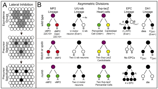
Asymmetric cell divisions generate sibling cells of distinct fates ('A', 'B') and constitute a fundamental mechanism that creates cell-type diversity in multicellular organisms. Antagonistic interactions between the Notch pathway and the intrinsic cell-fate determinant Numb appear to regulate asymmetric divisions in flies and vertebrates. During these divisions, productive Notch signaling requires sanpodo, which encodes a novel transmembrane protein. Here, we demonstrate that Drosophila sanpodo plays a dual role to regulate Notch signaling during asymmetric divisions - amplifying Notch signaling in the absence of Numb in the 'A' daughter cell and inhibiting Notch signaling in the presence of Numb in the 'B' daughter cell. In so doing, sanpodo ensures the asymmetry in Notch signaling levels necessary for the acquisition of distinct fates by the two daughter cells. These findings answer long-standing questions about the restricted ability of Numb and Sanpodo to inhibit and to promote, respectively, Notch signaling during asymmetric divisions.


Figures

Fig. 1.
Fig. 1.
Schematics of Notch-mediated lateral inhibition and the lineages of five pairs of sibling cells. (A) Initially all cells within three equivalence groups acquire the potential (gray) to adopt the precursor fate. One cell (gray) is singled out as the presumptive precursor from each cluster. This cell then acts through the Notch pathway to inhibit all other cells in the group (light gray) from adopting the precursor fate, such that individual precursors cells (black) segregate from each group (right). (B) Cell lineages of the five sibling pairs assayed in this paper shown for one hemisegment of wild type, Notch/spdo or numb mutant embryos. Lineages shown in color represent those in which we follow the fate of both sibling cells. Lineages shown in black/white are those in which we use eve expression (black) to follow the fate of one of the two sibling cells in each lineage.
Fig. 2.
Fig. 2.
sanpodo misexpression inhibits Notch signaling during lateral inhibition and wing development in a numb-dependent manner. (A) Four scutellar bristles arise in wild type. (B) Removing one copy of Notch has little effect on bristle number. (C-E) Expression of spdo by patched-GAL4 (o/e spdo) in the scutellar region promotes ectopic bristle formation (C), a phenotype enhanced by reducing Notch function 50% (D), and suppressed by co-expression of a numb-RNAi transgene (E) (Tang et al., 2005). (F) In wild type two sensory organ precursors, as labeled by anti-senseless (Nolo et al., 2000), develop in the scutellar region (arrows, brackets) of the late third instar wing imaginal disc. (G) spdo overexpression in this region promotes the formation of ectopic sensory organ precursors (arrow). (H) Average number of scutellar bristles per indicated genotype. Error bars indicate s.d. **P<10−4; ***P<10−10. (I) nubbin-GAL4-mediated expression of spdo in the wing (o/e spdo) leads to wing notching, vein thickening and reduced wing size. (J) Reduction of Notch function by 50% (N+/−; o/e spdo), using the Notch81k1 allele, enhanced these phenotypes. (K,L) Simultaneously reducing numb function by 50% (N+/−; numb+/−; o/e spdo, L) suppressed the phenotypes observed in N+/−; o/e spdo flies (K). Images in I-K are shown at identical magnification.
Fig. 3.
Fig. 3.
sanpodo exerts a context-dependent effect on Notch signaling during lateral inhibition. (A) Model of heart precursors and differentiated (mature) heart cells. Left, in a wild-type hemisegment two cardioblast precursors (red cells labeled ‘P’) each divide to produce two cardioblasts (red cells labeled ‘CB’), and two Svp-lacZ+ heart precursors (green cells labeled ‘P’) each divide to produce one Svp-lacZ+ pericardial cell (green, ‘A’ daughter cell) and one Svp-lacZ+ cardioblast (red, ‘B’ cell). Right, in numb mutant embryos heart precursors form normally but Svp-lacZ+ heart precursors produce only pericardial cells (green, ‘A’ cells). Precursors are shown for one hemisegment, whereas mature heart cells are shown for one full segment. (B-F) High-magnification views of two hemisegments of the dorsal mesoderm of embryos of indicated genotype labeled for Svp-lacZ+ (green) or Mef-2 (red), which labels mesodermal cells. (B-F′) High-magnification views of three segments of the heart of embryos of indicated genotype labeled for Svp-lacZ+ (green) and Nmr-1 (red), Nmr-1 specifically labels cardioblasts (Leal et al., 2009). (B) In wild-type stage 12 embryos, two Svp-lacZ+ heart precursors arise per hemisegment (arrows; dotted lines demarcate segment-sized regions in B-F). (B′) By stage 16, two rows of cardioblasts (red; yellow for Svp-lacZ+ cardioblasts) and Svp-lacZ+ pericardial cells (green) align on either side of the dorsal midline (thick white line). In B′-E′ one segment is bracketed and white lines indicate defined or inferred sibling relationships. (C) twist-GAL4-mediated expression of spdo in a wild-type embryo generates extra Svp-lacZ+ heart precursors (arrowhead) by stage 12, and extra Svp-lacZ+ pericardial cells and cardioblasts (arrowheads, C′) as well as extra Svp-lacZ cardioblasts (arrow, C′) by stage 16. (D) In numb mutant embryos two Svp-lacZ+ precursors arise normally (arrows), but each divides to produce two pericardial cells (green cells, D′), no Svp-lacZ+ cardioblasts develop in this background. (E) Expressing spdo throughout the mesoderm of a numb mutant embryo inhibits the formation of Svp-lacZ+ precursors (arrow), which leads to fewer Svp-lacZ+ pericardial cells (green cells, E′); note also the reduction in cardioblasts (red cells, E′). (F,F′) In wild-type mesodermal expression of a constitutively active form of Notch (NotchΔECN) leads to a near complete loss of Svp-lacZ+ heart precursors (arrowheads, F) and heart cells (F′). Anterior, left.
Fig. 4.
Fig. 4.
spdo potentiates Notch signaling in the absence of numb. Left and middle panels show lateral view; right panel shows dorsal view of eve expression in the mesoderm. (A-A″) In wild type, mesodermal Eve+ equivalence groups (arrows, A) generate Eve+ precursor cells (arrows, A′) that produce the EPCs (arrows, A″), and the Eve+ DA1 muscles (white arrows, A″). (B-B″) In numb mutant embryos, equivalence group formation and precursor selection occurs normally (arrows, B,B′); however, defects in asymmetric divisions lead to a doubling of EPC numbers (arrows, B″) and a loss of DA1 muscles (white arrows, B″). (C-C″) Eve+ equivalence groups form normally in numb mutant embryos that misexpress spdo in the mesoderm under the control of twist-GAL4 (arrows); however, Eve+ precursors fail to segregate from half of these groups (arrowhead, C′), resulting in fewer EPCs (arrows, C″). (D-D″) Eve+ equivalence groups form, albeit with reduced eve expression, upon mesodermal expression of a constitutively active form of Notch (NotchΔECN) in wild-type embryos; however, few precursors and EPCs arise in this background (arrows, D′,D″). Anterior, left.
Fig. 5.
Fig. 5.
numb regulates the subcellular localization of Spdo. Spdo localization (green) in ectodermal cells of otherwise wild-type (A) or numb mutant (B) embryos. (A) Spdo exhibits predominately diffuse and punctate localization in ectodermal cells of wild-type embryos. (B) Spdo exhibits increased localization to the cell membrane of ectodermal cells in numb mutant embryos. Each panel shows a single z-section of a stage 12 embryo taken at similar apical basal positions; both embryos were processed in parallel and imaged on a Leica SPII confocal microscope using identical parameters. Spdo misexpression mediated by scabrous-GAL4. Anterior, left.
Fig. 6.
Fig. 6.
spdo facilitates Numb-mediated inhibition of Notch signaling in the ‘B’ daughter cell during asymmetric divisions in the CNS. (A) In wild-type embryos vMP2 (arrows, ‘A’ cell) extends an axon anteriorly, while its sibling dMP2 (arrowheads, ‘B’ cell) expresses Odd (red) and extends an axon posteriorly. (B) In spdo mutant embryos both siblings adopt the Odd+ dMP2 fate (arrowheads) and extend axons posteriorly. (C) In numb mutant embryos both siblings adopt the vMP2 fate (arrows) and extend axons anteriorly. In C-F, an asterisk denotes the unrelated Odd+ MP1 neurons (red), which form in all genotypes shown but are obscured in some panels. (D) Single segment of wild-type embryo in which Notch has been overexpressed in the CNS. Left, daughter cells adopt normal vMP2 (arrows) and dMP2 (arrowheads) fates. Right, both daughter cells adopt the vMP2 fate and extend axons anteriorly (arrows). (E) Notch overexpression in spdo mutant embryos induces both daughter cells to adopt the vMP2 fate (arrows) and extend axons anteriorly in most hemisegments. (F) Fringe overexpression in spdo mutant embryos. Left: both daughter cells adopt the vMP2 fate (arrows) and extend axons anteriorly. Right: both daughter cells adopt the dMP2 fate (arrowheads) and extend axons posteriorly. (see Table S3 in the supplementary material). The dotted line in F indicates use of different focal planes from the same segment. Anterior is up; N>200 hemisegments per genotype. prospero-GAL4 was used for gene overexpression. See also Table S3 in the supplementary material.
Fig. 7.
Fig. 7.
Overexpression of Notch or Fringe bypasses the requirement for spdo function during EPC development. Each panel shows a lateral view of eve expression in the dorsal mesoderm. (A) Wild-type stage 14 embryo showing EPCs (arrows) and Eve+ DA1 muscles (arrowheads). (B) spdo mutant embryos exhibit a decrease in EPC number (arrows). (C) In numb mutant embryos EPC numbers double (arrows) and Eve+ DA1 muscles are lost (arrowheads). (D) twist-GAL4-mediated overexpression of Spdo (o/e spdo) in the mesoderm of wild-type embryos increased EPC numbers (arrows). (E,F) Identical experiments with either Notch (E) or Fringe (F) had little effect on EPCs (arrows) or DA1 muscles (arrowheads). (G-I) Mesodermal expression of spdo in a spdo mutant embryo rescued the spdo mutant phenotype (compare to B), whereas mesodermal overexpression of Notch (H) or Fringe (I) in spdo mutant embryos increased EPC numbers (arrows) and decreased DA1 muscle numbers (arrowheads), phenotypes similar to those of numb mutant embryos (compare with C). Numbers indicate average number of EPCs ±s.d. per bilateral side of an embryo. n≥20 embryo sides, except for o/e spdo; spdo (n=14). (J) Chart shows average number of EPCs per indicated genotype.
Fig. 8.
Fig. 8.
spdo facilitates Numb-mediated inhibition of Notch signaling during asymmetric divisions in the heart. (A) Wild-type pattern of sibling Svp-lacZ+ pericardial cells (green, ‘A’ cell) and cardioblasts (CBs; yellow, ‘B’ cell) in the stage 16 heart. In all panels one segment is bracketed, and white lines identify sibling relationships. (B) In spdo mutant embryos both Svp-lacZ+ daughter cells normally adopt the cardioblast or ‘B’ fate (yellow, arrowheads). (C) In numb mutant embryos both daughter cells adopt the pericardial or ‘A’ fate (green, arrowheads). (D,E) twist-GAL4-mediated expression of GFP (D), or Neuralized and Delta (E), in spdo mutant embryos causes both daughter cells to adopt the ‘A’ fate at low frequency (arrowhead), and increases the frequency of both daughter cells adopting alternate fates (‘A/B’). Note GFP overexpression has no effect on any other lineage tested (Fig. 3, see Tables S2 and S3 in the supplementary material). (F) Notch overexpression in wild type induces both daughter cells to adopt the ‘A’ fate at low frequency (arrowhead). (G) Notch overexpression in spdo mutant embryos increases the frequency at which both daughter cells adopt the ‘A’ fate (arrowheads). (H) Fringe overexpression in wild type directs both daughter cells to adopt the ‘A’ fate most of the time (arrowheads). (I) Fringe overexpression in spdo mutant embryos directs both daughter cells to adopt the ‘A’ fate (green, arrowheads) essentially all the time (arrowheads). Anterior, left. Quantification of sibling fates. n>800 sibling pairs assayed per genotype, except for numb (n=200). See also Table S3 in the supplementary material. (J) Model of spdo function during asymmetric divisions. Top, in spdo mutant embryos Notch signaling activity remains below the threshold level (dotted line) required to induce the ‘A’ fate, and both daughter cells adopt the ‘B’ fate. Bottom, in the absence of Numb, Spdo amplifies Notch signaling activity above the threshold required to induce the ‘A’ fate. In the presence of Numb, Spdo facilitates the ability of Numb to inhibit Notch signaling ‘B’ daughter cell, thereby reducing signaling activity below that observed in the absence of spdo.

Similar articles

Cited by

References

    1. Ahmed A., Chandra S., Magarinos M., Vaessin H. (2003). Echinoid mutants exhibit neurogenic phenotypes and show synergistic interactions with the Notch signaling pathway. Development 130, 6295-6304 - PubMed
    1. Berdnik D., Torok T., Gonzalez-Gaitan M., Knoblich J. A. (2002). The endocytic protein alpha-Adaptin is required for numb-mediated asymmetric cell division in Drosophila. Dev. Cell 3, 221-231 - PubMed
    1. Blair S. S. (2000). Notch signaling: Fringe really is a glycosyltransferase. Curr. Biol. 10, R608-R612 - PubMed
    1. Brand A. H., Perrimon N. (1993). Targeted gene expression as a means of altering cell fates and generating dominant phenotypes. Development 118, 401-415 - PubMed
    1. Calleja M., Moreno E., Pelaz S., Morata G. (1996). Visualization of gene expression in living adult Drosophila. Science 274, 252-255 - PubMed

Publication types

MeSH terms