. 2009 Dec;136(23):3959-67.
doi: 10.1242/dev.039768.

Temporal and spatial dissection of Shh signaling in genital tubercle development

Affiliations

Temporal and spatial dissection of Shh signaling in genital tubercle development

Congxing Lin et al. Development. 2009 Dec.

Abstract

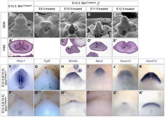
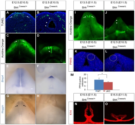
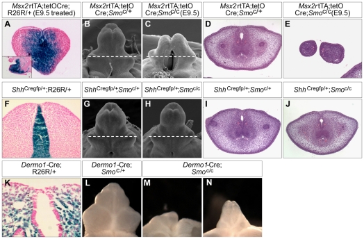
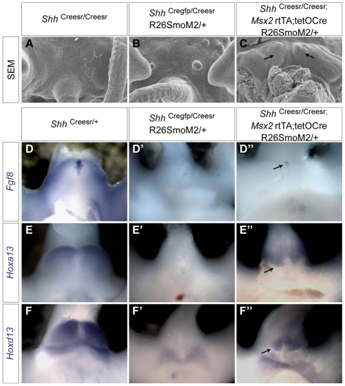
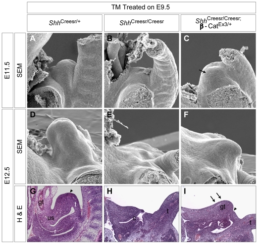
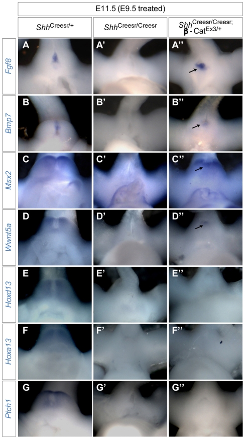
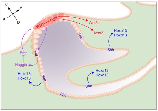
Genital tubercle (GT) initiation and outgrowth involve coordinated morphogenesis of surface ectoderm, cloacal mesoderm and hindgut endoderm. GT development appears to mirror that of the limb. Although Shh is essential for the development of both appendages, its role in GT development is much less clear than in the limb. Here, by removing Shh at different stages during GT development in mice, we demonstrate a continuous requirement for Shh in GT initiation and subsequent androgen-independent GT growth. Moreover, we investigated the Hh responsiveness of different tissue layers by removing or activating its signal transducer Smo with tissue-specific Cre lines, and established GT mesenchyme as the primary target tissue of Shh signaling. Lastly, we showed that Shh is required for the maintenance of the GT signaling center distal urethral epithelium (dUE). By restoring Wnt-Fgf8 signaling in Shh(-/-) cloacal endoderm genetically, we revealed that Shh relays its signal partly through the dUE, but regulates Hoxa13 and Hoxd13 expression independently of dUE signaling. Altogether, we propose that Shh plays a central role in GT development by simultaneously regulating patterning of the cloacal field and supporting an outgrowth signal.


Figures

Fig. 1.
Fig. 1.
Phenotypic and gene expression analysis of Shh conditional knockout GTs. (A-E) Scanning electron microscopy (SEM) analyses of E15.5 male genital tubercles (GTs) of control (A) and Shh-cKO (B-E) mice. The timing of Tamoxifen (Tm) treatment is indicated. Proximal hypospadias is indicated by arrows in B-E. (A′,C-E′). Hematoxylin and Eosin (H&E)-stained cross-sections of wild-type (A′) and late Shh-cKO (C′-E′) GTs. Urethral epithelium and mesenchymal condensations are indicated by arrowheads and arrows, respectively. (F-K′) Whole-mount in situ analysis on E12.5 GTs from Shh-cKOs (F′-K′) and littermate controls (F-K). All embryos were exposed to Tm at E10.5.
Fig. 2.
Fig. 2.
Cell death and proliferation analyses in Shh-cKOs. (A,B) TUNEL analysis on coronal sections of GTs (Tm treatment at E10.5) showing distal mesenchymal apoptosis in control (A, arrows) and excessive urethral epithelium (UE) staining in Shh-cKO (B, arrowheads) GT. (C,D) Whole-mount Acridine Orange (AO) staining showing patterned mesenchymal cell death in control (C, arrows), and a drastically augmented UE staining in Shh-cKOs (D, arrowheads). (E-H) Whole-mount in situ hybridization on E12.5 GTs from Shh-cKOs (F,H) and littermate controls (E,G) using the probes indicated. Bmp4 expression is normally detected in the genital mesenchyme (E), and is increased in the distal GT of the mutant (F). Nog is expressed in the distal mesenchyme (arrows in G), and is undetectable in the mutant (H). (I,J) AO staining of E13.5 control (I) and Shh-cKO (J) GT (both were treated with Tm at E11.5) showing a normal mesenchymal staining pattern in the control (I, arrows), and a UE-restricted staining in the Shh-cKO (J, arrowheads). (K-M) Phospho-histone H3 (PHH3) staining on coronal sections of Shh-cKO (L) and controls (K) showing a 20% reduction (M) in PHH3-positive cells in the yellow-circled region. n=8, *P=0.0027. (N,O) Indirect immunofluorescence using a K14 antibody on E15.5 control (N) and Shh-cKO (O) male GT (TM treated at E11.5).
Fig. 3.
Fig. 3.
Phenotypic analyses of conditional knockout Smo mutant GTs. (A) β-gal staining on cross-section or sagittal section (inset) of an E12.5 Msx2-rtTA;tetO-Cre;R26R/+ GT (Dox treatment at E9.5) showing strong Cre expression in UE and mesenchyme, especially the dorsal GT mesenchyme. (B,C) SEM analysis of E15.5 male UEMes-Smo-cKO (C) and littermate control (B) GT. (D,E) H&E staining of UEMes-Smo-cKO (E) and littermate control (D) GT. The planes of sections are indicated in B and C (dashed lines). (F) β-gal staining on cross-sections of a E12.5 ShhCregfp/+;R26R/+ GT demonstrating Cre expression in UE. (G-J) SEM and histological analyses of UE-Smo-cKOs (H,J) and littermate controls (G,I). (K) β-gal staining on cross-sections of a E10.5 Dermo1-Cre;R26R/+ GT showing that the expression was exclusively in the cloacal mesenchyme. (L-N) Light microscopy of Mes-Smo-cKOs (M,N) and control (L). GTs from E15.5 male mouse embryos were analyzed in B-E,G-J,L-N.
Fig. 4.
Fig. 4.
Rescuing GT agenesis in Shh-/- embryos by restoring Hh responsiveness. (A-C) SEM analysis of E12.5 genital region of Shh-/- mouse embryos (A) and Shh-/- embryos with Hh responsiveness restored in UE (B) or in UE and mesenchyme together (C). Bilateral growth in the UEmes-rescued line is indicated by arrows in C. (D-F″) Whole-mount in situ hybridization on E11.5 UE-rescued Shh-/- (D′,E′,F′), UEmes-rescued Shh-/- (D″,E″,F″) and ShhCreesr/+ littermate control (D,E,F) GT. The rescued expression of Fgf8, Hoxa13 and Hoxd13 is indicated by arrows.
Fig. 5.
Fig. 5.
Phenotypic analysis of dUE-rescued Shh-/- GTs. (A-C) SEM analysis of E11.5 control (A), Shh-/- (B) and dUE-rescued Shh-/- (C) mouse embryos showing agenesis of the GT in Shh-/- (B) and prominent GT growth in the rescued Shh-/- (arrow in C). (D-F) SEM analysis of E12.5 embryos of the genotypes indicated showing that the rescued GT (F) was much smaller than in the wild type (D). (G-I) H&E staining on mid-sagittal sections of E12.5 embryos showing a considerable dorsal growth (arrows in I) and a persistent cloaca in the dUE-rescued Shh-/-. The distal end of the UE is indicated by arrowheads in G,I. gt, genital tubercle; us, urogenital sinus; r, rectum; c, cloaca; t, tail.
Fig. 6.
Fig. 6.
Gene expression analysis of dUE-rescued Shh-/- GTs. (A-G″) Mouse embryos were treated with Tm at E9.5 and collected on E11.5 for in situ hybridization with the probes indicated. The expected expression pattern of Fgf8, Bmp7, Msx2 and Wnt5a was observed in the controls (A-D), was absent in Shh-/- GTs (A′-D′), but was fully (A″,B″, arrows) or partially (C″,D″, arrows) restored in dUE-rescued Shh-/- GTs. Expression of Hoxd13 and Hoxa13 was either severely reduced (E′,F′) or undetectable (E″,F″) in both Shh-/- and dUE-rescued Shh-/- GTs. Ptch1 expression was not detected in either Shh-/- or dUE-rescued GTs (G-G″).
Fig. 7.
Fig. 7.
Coordination of Shh and dUE signaling in patterning the cloaca and in formation of the mouse GT. Shh signals (dark-blue arrows) to adjacent cloacal mesenchyme to activate Hox gene expression and to coordinate patterning and growth of the whole cloacal field. The addition of dUE signaling (red), activated by Wnt signaling, functions as an AER-like outgrowth genetic cassette, triggering the distal growth of the mesenchymal cells and resulting in the formation of the genital primordia (red arrows). Shh might also signal to genital mesenchyme to activate noggin expression, which in turn inhibits Bmp activity and thus maintains dUE signaling (purple arrows).

Similar articles

Cited by

References

    1. Allard P., Tabin C. J. (2009). Achieving bilateral symmetry during vertebrate limb development. Semin. Cell Dev. Biol. 20, 479-484 - PubMed
    1. Barnes E. A., Kong M., Ollendorff V., Donoghue D. J. (2001). Patched1 interacts with cyclin B1 to regulate cell cycle progression. EMBO J. 20, 2214-2223 - PMC - PubMed
    1. Cohn M. J. (2004). Developmental genetics of the external genitalia. Adv. Exp. Med. Biol. 545, 149-157 - PubMed
    1. Echelard Y., Epstein D. J., St-Jacques B., Shen L., Mohler J., McMahon J. A., McMahon A. P. (1993). Sonic hedgehog, a member of a family of putative signaling molecules, is implicated in the regulation of CNS polarity. Cell 75, 1417-1430 - PubMed
    1. Freitas R., Zhang G., Cohn M. J. (2007). Biphasic Hoxd gene expression in shark paired fins reveals an ancient origin of the distal limb domain. PLoS ONE 2, e754 - PMC - PubMed

Publication types

MeSH terms