. 2009 Dec;136(23):3969-78.
doi: 10.1242/dev.039438.

Dosage-dependent hedgehog signals integrated with Wnt/beta-catenin signaling regulate external genitalia formation as an appendicular program

Affiliations

Dosage-dependent hedgehog signals integrated with Wnt/beta-catenin signaling regulate external genitalia formation as an appendicular program

Shinichi Miyagawa et al. Development. 2009 Dec.

Abstract

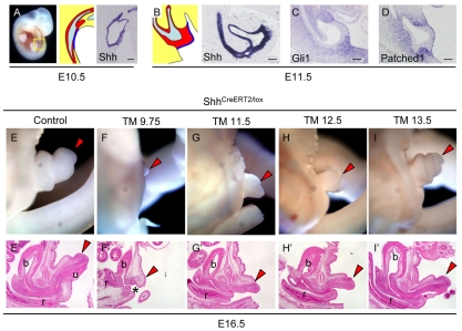
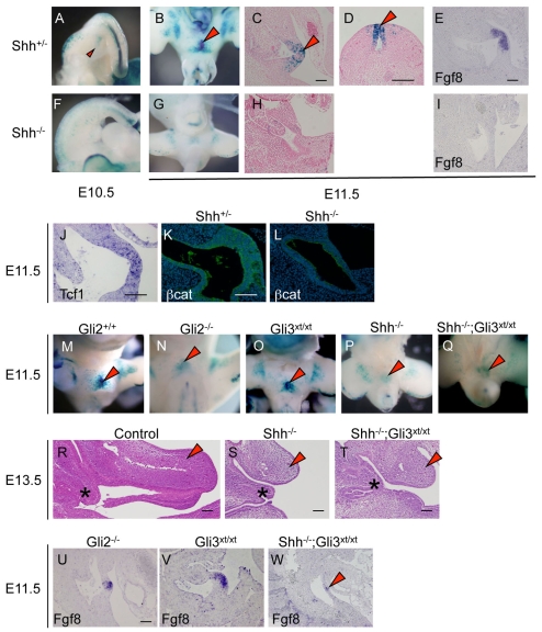
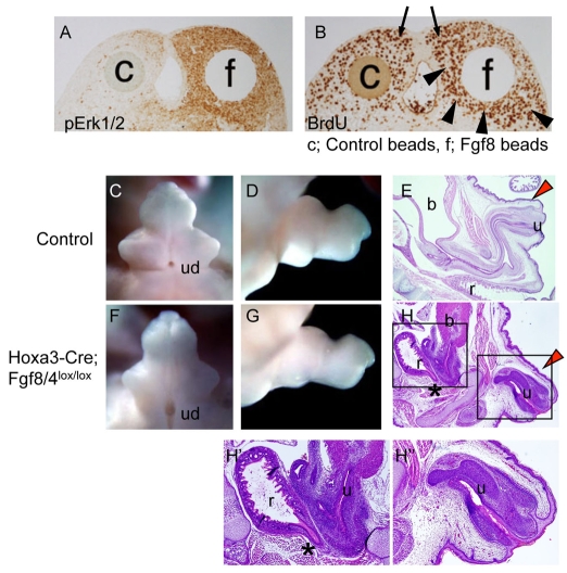
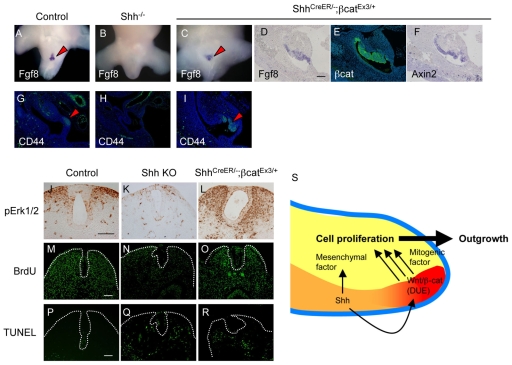
Embryonic appendicular structures, such as the limb buds and the developing external genitalia, are suitable models with which to analyze the reciprocal interactions of growth factors in the regulation of outgrowth. Although several studies have evaluated the individual functions of different growth factors in appendicular growth, the coordinated function and integration of input from multiple signaling cascades is poorly understood. We demonstrate that a novel signaling cascade governs formation of the embryonic external genitalia [genital tubercle (GT)]. We show that the dosage of Shh signal is tightly associated with subsequent levels of Wnt/beta-catenin activity and the extent of external genitalia outgrowth. In Shh-null mouse embryos, both expression of Wnt ligands and Wnt/beta-catenin signaling activity are downregulated. beta-catenin gain-of-function mutation rescues defective GT outgrowth and Fgf8 expression in Shh-null embryos. These data indicate that Wnt/beta-catenin signaling in the distal urethral epithelium acts downstream of Shh signaling during GT outgrowth. The current data also suggest that Wnt/beta-catenin regulates Fgf8 expression via Lef/Tcf binding sites in a 3' conserved enhancer. Fgf8 induces phosphorylation of Erk1/2 and cell proliferation in the GT mesenchyme in vitro, yet Fgf4/8 compound-mutant phenotypes indicate dispensable functions of Fgf4/8 and the possibility of redundancy among multiple Fgfs in GT development. Our results provide new insights into the integration of growth factor signaling in the appendicular developmental programs that regulate external genitalia development.


Figures

Fig. 1.
Fig. 1.
Temporal requirements for hedgehog signaling during GT initiation and outgrowth. (A,B) Murine GT development and Shh expression at E10.5 (A) and E11.5 (B). Red regions indicate Shh-expressing endoderm-derived epithelia. The cloacal region includes the thick endodermal epithelia (in red, B). The dark-blue line indicates adjacent ectoderm-derived epithelium. Cloacal cavity is in light blue. (C,D) Gli1 (C) and patched 1 (D) expression in GT mesenchyme and ectoderm at E11.5. (E-I′) Phenotypes after temporally staggered ablation of Shh. GT phenotypes of ShhCreERT2/lox embryos treated with TM at E9.75 (F), E11.5 (G), E12.5 (H) and E13.5 (I) versus the control GT (no TM administration, E). TM administration at E9.75 induced severe GT aplasia with a persistent cloaca (asterisk). Arrowheads indicate GTs or GT rudiments. b, bladder; r, rectum; u, urethra. Scale bars: 100 μm.
Fig. 2.
Fig. 2.
Expression of Wnt ligands in control and Shh KO mouse embryos at E11.5. (A-H) Ectodermal expression (arrowheads) of Wnt3 (A,E), Wnt4 (B,F), Wnt7a (C,G) and Wnt9b (D,H) is decreased in Shh KO embryos. Scale bar: 100 μm.
Fig. 3.
Fig. 3.
Wnt/β-catenin signaling is responsive to hedgehog signaling dosage in the mouse GT. (A-E) TopGAL activity (arrowheads) is detectable in the distal region of the GT, including the DUE at E10.5 (A) and E11.5 (B-D). lacZ staining (C,D, arrowheads) overlaps with the Fgf8 expression domain (E). (F-I) TopGAL activity (F-H) and Fgf8 expression (I) are not detected in Shh KO embryos. (J) Tcf1 is expressed in the distal GT. (K,L) β-catenin protein localizes predominantly to the endodermal epithelium (K, green) and decreases in Shh KO embryos (L). (M-Q) Gli2 KO embryos have decreased TopGAL reporter activity (N) relative to control (M) and Gli3xt/xt embryos (O). Shh-/-;Gli3xt/xt double mutants exhibit weak, but detectable, TopGAL activity (Q, arrowhead) as compared with Shh KO embryos (P). (R-T) Sections of control (R), Shh KO (S) and Shh-/-;Gli3xt/xt (T) embryos. GT outgrowth is restored in the Shh-/-;Gli3xt/xt double mutants at E13.5 (arrowhead). Asterisk indicates the distal end of the urorectal septum. (U-W) GT outgrowth and Fgf8 expression in hedgehog pathway mutants at E11.5. Decreased Fgf8 expression and GT hypoplasia in Gli2 KO embryos (U, compare with E). Gli3xt/xt embryos have normal GT protrusion and Fgf8 expression (V). Shh-/-;Gli3xt/xt embryos exhibit a small bud structure with some Fgf8 expression (W, arrowhead). Scale bars: 100 μm.
Fig. 4.
Fig. 4.
Gene expression in β-catenin loss-of-function mutants. (A) Isl1-Cre-mediated lacZ expression in R26R mice. Cre is active in the caudal E9.0 embryo (arrowhead). (B-E) E11.5 control embryos (B,C). Fgf8 (D) and Shh (E) expression (arrowheads) in IslCre/+;Ctnnb1lox/lox embryos. (F-K) Failed GT outgrowth in E11.5 endoderm-specific β-catenin (βcat) conditional mutants (F,G). Green, immunofluorescence for β-catenin; blue, Hoechst counterstain. Fgf8 expression is abnormal (H,J), whereas Gli1 is normal (I,K), at E11.5. (L) ChIP/PCR on GT and limb reveals that β-catenin can bind to the murine CR3 region but not to the 5′ flanking region. Acetylated histone H3 is bound to both regions. (M) The CR3 putative enhancer activates expression of a luciferase reporter in response to overexpressed wild-type or constitutively active β-catenin. *, P<0.05 versus empty vector control. Scale bar: 100 μm.
Fig. 5.
Fig. 5.
Fgf8 and Fgf4 are dispensable for mouse GT development. (A,B) Anti-pErk1/2 and BrdU immunostaining in GT explants. pErk1/2 activity is restricted to distal GT with the control (c) bead (A). pErk1/2 (A) and BrdU incorporation (B) are augmented in regions adjacent to Fgf8 (f) beads. Arrowheads and arrows indicate augmented and endogenous cell proliferation, respectively. (C-H″) GT phenotype of control (C-E) and Hoxa3-Cre;Fgf8/4lox/lox mutant embryos (F-H″) at E14.5 (C,D,F,G) and E18.5 (E,H-H″). GT outgrowth appears normal in mutants (arrowheads), but the urogenital duct (ud) is wide open and anal stenosis (asterisk) and urethral narrowing with proximal atresia are also present. The distal urethra appears normal. b, bladder; r, rectum; u, urethra.
Fig. 6.
Fig. 6.
Rescue of GT outgrowth in Shh-null mutants by constitutively active β-catenin. (A-J) Control GTs (A,F) and Shh KO mouse embryos with GT agenesis (B,G,I). ShhCreERT2/-;Ctnnb1Ex3/+ embryos have GT protrusion at E11.5 and continued GT outgrowth with preputial fold at E13.5 (C,H). Further outgrowth is evident by E18.5 (J). Gli1 expression is undetectable in ShhCreERT2/-;Ctnnb1Ex3/+ embryos at E11.5 (D,E). Arrowheads indicate GT or prospective GT regions.
Fig. 7.
Fig. 7.
Regulation of Fgf8 expression by Shh and β-catenin. (A-I) Fgf8 (A,B) and Cd44 (G,H) expression (arrowheads) in control (A,G) versus Shh KO (B,H) DUE. Expression is recovered in ShhCreERT2/-;Ctnnb1Ex3/+ embryos (arrows in C,I). Fgf8 and Cd44 expression overlap with regions of augmented β-catenin and Axin2 (D-F,I). (J-L) pErk1/2 staining in control (J) versus Shh KO (K) and ShhCreERT2/-;Ctnnb1Ex3/+ (L) embryos. (M-R) BrdU labeling (M-O) and TUNEL assays (P-R). Shh KO embryos show significantly decreased cell proliferation (N). By contrast, expression of stabilized β-catenin in Shh KO mutants restores cell proliferation (O). Mesenchymal apoptosis was unchanged in ShhCreERT2/-;Ctnnb1Ex3/+ embryos as compared with Shh KO embryos (Q,R). Dotted lines in the midline GT indicate the basal layer of the endodermal and ectodermal epithelia. (S) Proposed signaling cascade for GT outgrowth. Shh signaling is required for GT initiation and subsequent outgrowth. Shh is expressed moderately in the DUE (red) and strongly in urethral epithelium (orange). Wnt ligands are expressed in the distal GT ectoderm, and Wnt/β-catenin activity is detected in the distal region of the GT, including the DUE. Wnt/β-catenin activity depends on Shh signaling. This Wnt/β-catenin activity, and possibly urethral plate-derived Shh, induce production of mitogenic factors leading to GT outgrowth. Yellow, mesenchyme; blue line, ectoderm-derived epithelium. Scale bars: 100 μm.

Similar articles

Cited by

References

    1. Ahn K., Mishina Y., Hanks M. C., Behringer R. R., Crenshaw E. B., 3rd (2001). BMPR-IA signaling is required for the formation of the apical ectodermal ridge and dorsal-ventral patterning of the limb. Development 128, 4449-4461 - PubMed
    1. Araki K., Imaizumi T., Okuyama K., Oike Y., Yamamura K. (1997). Efficiency of recombination by Cre transient expression in embryonic stem cells: comparison of various promoters. J. Biochem. 122, 977-982 - PubMed
    1. Barrow J. R., Thomas K. R., Boussadia-Zahui O., Moore R., Kemler R., Capecchi M. R., McMahon A. P. (2003). Ectodermal Wnt3/beta-catenin signaling is required for the establishment and maintenance of the apical ectodermal ridge. Genes Dev. 17, 394-409 - PMC - PubMed
    1. Beermann F., Kaloulis K., Hofmann D., Murisier F., Bucher P., Trumpp A. (2006). Identification of evolutionarily conserved regulatory elements in the mouse Fgf8 locus. Genesis 44, 1-6 - PubMed
    1. Benazet J. D., Bischofberger M., Tiecke E., Goncalves A., Martin J. F., Zuniga A., Naef F., Zeller R. (2009). A self-regulatory system of interlinked signaling feedback loops controls mouse limb patterning. Science 323, 1050-1053 - PubMed

Publication types

MeSH terms