. 2009 Dec;136(23):3979-89.
doi: 10.1242/dev.037267.

Semaphorin3a regulates endothelial cell number and podocyte differentiation during glomerular development

Affiliations

Semaphorin3a regulates endothelial cell number and podocyte differentiation during glomerular development

Kimberly J Reidy et al. Development. 2009 Dec.

Abstract

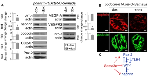
Semaphorin3a (Sema3a), a chemorepellant guidance protein, plays crucial roles in neural, cardiac and peripheral vascular patterning. Sema3a is expressed in the developing nephron, mature podocytes and collecting tubules. Sema3a acts as a negative regulator of ureteric bud branching, but its function in glomerular development has not been examined. Here we tested the hypothesis that Sema3a regulates glomerular vascular development using loss- and gain-of-function mouse models. Sema3a deletion resulted in defects in renal vascular patterning, excess endothelial cells within glomerular capillaries, effaced podocytes with extremely wide foot processes and albuminuria. Podocyte Sema3a overexpression during organogenesis resulted in glomerular hypoplasia, characterized by glomerular endothelial cell apoptosis, delayed and abnormal podocyte foot process development, a complete absence of slit diaphragms and congenital proteinuria. Nephrin, WT1 and VEGFR2 were downregulated in Sema3a-overexpressing kidneys. We conclude that Sema3a is an essential negative regulator of endothelial cell survival in developing glomeruli and plays a crucial role in podocyte differentiation in vivo. Hence, a tight regulation of Sema3a dosage is required for the establishment of a normal glomerular filtration barrier.


Figures

Fig. 1.
Fig. 1.
Sema3a deletion results in excess glomerular endothelial cells and podocyte foot process effacement. (A) PAS-stained wild-type glomerulus (+/+) with open capillaries; Sema3a-null (-/-) glomerulus shows multiple blue nuclei in the capillary loops and few open lumina. (B) TEM: newborn Sema3a+/+ (3A+/+) glomeruli demonstrate intact foot processes (fp), slit diaphragms (SDs), GBM and endothelial cells (EC). Sema3a-/- (3A-/-) glomeruli show multiple EC within the capillary loop, wide foot processes (efp: effaced foot process, red arrowheads) joined by occluding-junctions (OJ). P, podocyte; Cap, capillary lumen. (C) TEM morphometric analysis: the number of EC per capillary loop is two-fold higher, the foot processes are ~seven-fold wider, and the SD density is five-fold lower in Sema3a-/- glomeruli versus wild-type glomeruli (n=8 mice per group, P=0.0002). *, P<0.05. (D) Western blot showing albuminuria in Sema3a-/- newborn mice. Scale bars: in A, 20 μm; in B, 1 μm for upper panel, 200 nm in lower panel.
Fig. 2.
Fig. 2.
Sema3a deletion causes excess renal endothelial cells and disrupts vascular patterning. (A) β-Gal-stained kidney sections (80 μm thick) from newborn progeny of Flk1/lacZ+/-: Sema3a+/- mice. In wild-type (3A+/+) kidneys, most Flk1-positive endothelial cells localize to the cortex; Sema3a-/- (3A-/-) kidneys show abnormal renal vascular patterning, and increased endothelial cells can be observed throughout. (B) Newborn kidneys with endothelial cells immunostained for CD31 (red) and proliferating cells for PCNA (green) showing that most proliferation localizes to tubules (T) in both Sema3a+/+ and Sema3a-/- newborn mice. Insets: higher magnification of glomeruli (G) with endothelial cell proliferation (yellow) exclusively in Sema3a-/- mice. (C,D) Quantification of CD31 immunostaining confirming increased cortical and glomerular endothelial cells in Sema3a-/- kidneys: (C) proportion of CD31-positive cortical area (%); (D) proportion of CD31-positive glomerular area (%). *, P<0.05. Scale bars: in A, 200 μm for upper panel, 50 μm for lower panel; in B, 25 μm.
Fig. 3.
Fig. 3.
Loss of Sema3a promotes glomerular endothelial survival and migration. (A,B) Glomerular apoptosis detected by TUNEL assay (A) and by cleaved caspase 3 immunofluorescence (B) is shown; insets show apoptotic nuclei at higher magnification, nuclei are labeled with DAPI (blue). Merge between WT1 and DAPI appears pink, demonstrating nuclear WT1 staining. (A) In Sema3a+/+ glomeruli, apoptotic nuclei (green) co-localize with Griffonia simplicifolia Lectin I-labeled endothelium (red), indicating endothelial cell apoptosis (yellow in merged images), whereas no apoptotic endothelial cells were detected in the representative Sema3a-/- glomerulus shown. (B) Apoptotic cells were rare in both Sema3a-/- and Sema3a+/+ glomeruli, most Sema3a+/+ apoptotic nuclei (green) were not WT1-positive podocytes, and most Sema3a-/- apoptotic nuclei (green) were WT1-positive podocytes (yellow in merge). (C) Quantitative migration assay showing that Sema3a decreases mouse endothelial cell migration (MGEC) by approximately 50%. Data are expressed as mean ± s.e.m. of four experiments. *, P<0.05. (D,E) Sema3a blunts embryonic kidney chemo-attraction for MGEC. (D) Co-culture of embryonic kidneys with MGEC showing MGEC migrating towards the explant (control). (E) Co-culture of embryonic kidneys with MGEC exposed to Sema3a (250 ng/ml) prevented MGEC migration towards the explant (Sema3A). Scale bars: in A, 10 μm; in B, 10 μm; in D,E, 50 μm.
Fig. 4.
Fig. 4.
Pax2 is downregulated in Sema3a-/- kidneys. Western blots showing Pax2 downregulation in Sema3a-/- versus Sema3a+/+ kidneys. Nephrin, podocin, WT1, VEGFA, VEGFR2 and neuropilin 1 (nrp-1) expression was similar in Sema3a-/- and wild-type kidneys. Representative immunoblots of pooled samples (n=5 mice per genotype) are shown; bar graphs show densitometric analysis, expressed as fold change ± s.e.m. (n≥3), Sema3a-/- versus Sema3a+/+, corrected for actin. *, P<0.05.
Fig. 5.
Fig. 5.
Overexpression of podocyte Sema3a results in glomerular hypoplasia, undifferentiated podocytes and congenital proteinuria. (A) qPCR showing four-fold upregulation of Sema3a mRNA in isolated glomeruli of adult mice overexpressing Sema3a (gray bar), after a one-week induction with doxycycline versus controls (white bar, uninduced podocin-rtTA: tet-O-Sema3a and tet-O-Sema3a or podocin-rtTA on doxycyline). Data were normalized to ubiquitin and expressed as a fold-change from controls (n=4 per group, *, P<0.05). (B,C) PAS staining (B) demonstrating smaller glomeruli in newborn Sema3a-overexpressing (+dox) mice induced during kidney organogenesis. Glomerular volume (C) is decreased in Sema3a-overexpressing mice (gray bars) versus controls (white bars), n=4 per group. *, P<0.05. (D) TEM images demonstrating immature cuboidal podocytes (P) linked by occluding junctions (OJ) instead of slit diaphragms (SD) in newborn Sema3a-overexpressing mice induced during organogenesis (+dox). The endothelial cell (EC) appears swollen (red double-headed arrows) in induced glomeruli. Uninduced age-matched mice (-dox) and single transgenic littermates exhibit intact podocyte foot processes (fp) with normal GBM and fenestrated endothelium. Cap: capillary lumen, efp: effaced foot process (red arrowheads). (E) TEM morphometric analysis showing significantly wider foot processes (effacement), slit diaphragm absence and increase in occluding-junction number in Sema3a-overexpressing kidneys. *, P<0.05. (F) Western blot showing albuminuria in Sema3a-overexpressing mice (+dox) vs controls (-dox). Scale bars: in B, 10 μm; in D, 1 μm for upper panel, 500 nm for lower panel.
Fig. 6.
Fig. 6.
Podocyte foot process development is delayed and abnormal in mice overexpressing podocyte Sema3a. (A,B) TEM: Sema3a-overexpressing (+dox) glomeruli at completion of nephrogenesis (2 weeks) have wide podocyte foot processes (arrowheads) and absent slit diaphragms; a morphometric analysis is shown in B. Note that the endothelial cells (EC) are swollen (double-headed arrow). Controls (-dox) exhibit intact podocyte foot processes (fp), normal GBM and fenestrated endothelium. Cap: capillary lumen. (C) Urine albumin immunoblot demonstrates albuminuria in 2-week-old induced Sema3a-overexpressing mice (+dox), which is absent in controls (-dox). Scale bars: 1 μm.
Fig. 7.
Fig. 7.
Podocyte Sema3a overexpression downregulates nephrin, WT1 and VEGFR2. (A) Immunoblots showing nephrin, WT1 and VEGFR2 downregulation in Sema3a-overexpressing mice (+dox E12 to 2 weeks of age). Representative blots and densitometric analysis of three separate experiments are shown; data are expressed as fold change ± s.e.m. after correction for actin; +dox vs -dox were compared using pooled samples of n=13-20 mice per group; *, P<0.05. (B) Immunostaining demonstrating that nephrin expression is decreased in newborn Sema3a-overexpressing glomeruli (+dox), whereas podocin expression is unchanged. (C) Proposed model of the Sema3a effect on PAX2 and WT1 signaling. Previously described pathways are shown in blue: PAX2 is capable of either upregulation or repression of WT1 expression, depending upon the absence or presence, respectively, of the transcriptional co-repressor groucho TLE4. WT1 stimulates nephrin expression, and it downregulates PAX2 through a negative-feedback loop. The effects of Sema3a on these pathways are shown in red: Sema3a negatively regulates WT1 and nephrin and might positively regulate PAX2. Therefore, upon Sema3a deletion, PAX2 expression is downregulated, and, upon Sema3a overexpression, WT1 and nephrin expression is decreased.
Fig. 8.
Fig. 8.
Podocyte Sema3a overexpression negatively regulates glomerular endothelial cell survival, and podocyte number is not changed. (A) Several apoptotic nuclei (brown) identified by TUNEL in Sema3a- overexpressing glomeruli (+dox) versus virtually none in controls (-dox). (B) Quantification showing an eight-fold increase in apoptotic nuclei per glomerulus in Sema3a-overexpressing mice (gray bar) versus controls (white bar). *, P<0.05. (C,D) Identification of apoptotic cell type is shown by TUNEL assay and EC labeling (C) or by activated-caspase-3 immunofluorescence and podocyte labeling (D); nuclei are labeled with DAPI (blue). (C) In Sema3a-overexpressing glomeruli (+dox), apoptotic nuclei (green) co-localize with Griffonia simplicifolia Lectin I-labeled endothelium (red), indicating endothelial cell apoptosis (yellow in merged images; insets show apoptotic nuclei at a higher magnification). Two representative glomeruli are shown. (D) WT1 (red) and activated caspase 3 (green) immunostaining. Control glomeruli (-dox) show WT1-positive podocytes and no apoptosis (negative for activated caspase 3). Two representative glomeruli from Sema3a-overexpressing mice (+dox) are shown. In most glomeruli, apoptotic nuclei are WT1-negative (top inset), indicating that the apoptotic cells are not podocytes. Only 10% of apoptotic cells were deemed to be podocytes (WT1-positive and caspase-3-positive, shown in yellow, bottom inset). (E) Podocyte number quantification (WT1-positive nuclei per mm2 glomerular area) showing similar podocyte numbers in controls (white bar) and Sema3a-overexpressing mice (gray bar). n=40-100 glomeruli per group; P=ns. Scale bars: in C, 5 μm; in D, 10 μm.

Similar articles

Cited by

References

    1. Abrahamson D. R., Robert B., Hyink D. P., St John P. L., Daniel T. O. (1998). Origins and formation of microvasculature in the developing kidney. Kidney Int. Suppl. 67, S7-S11 - PubMed
    1. Avner E. D., Piesco N. P., Sweeney W. E., Jr, Ellis D. (1988). Renal epithelial development in organotypic culture. Pediatr. Nephrol. 2, 92-99 - PubMed
    1. Bagnard D., Vaillant C., Khuth S. T., Dufay N., Lohrum M., Puschel A. W., Belin M. F., Bolz J., Thomasset N. (2001). Semaphorin 3A-vascular endothelial growth factor-165 balance mediates migration and apoptosis of neural progenitor cells by the recruitment of shared receptor. J. Neurosci. 21, 3332-3341 - PMC - PubMed
    1. Behar O., Golden J. A., Mashimo H., Schoen F. J., Fishman M. C. (1996). Semaphorin III is needed for normal patterning and growth of nerves, bones and heart. Nature 383, 525-528 - PubMed
    1. Cai Y., Brophy P., Levitan I., Stifani S., Dressler G. (2003). Groucho suppresses Pax2 transactivation by inhibition of JNK-mediated phosphorylation. EMBO J. 22, 5522-5529 - PMC - PubMed

Publication types

MeSH terms