. 2010 Feb;84(3):1641-7.
doi: 10.1128/JVI.01979-09. Epub 2009 Nov 11.

NS1' of flaviviruses in the Japanese encephalitis virus serogroup is a product of ribosomal frameshifting and plays a role in viral neuroinvasiveness

Affiliations

NS1' of flaviviruses in the Japanese encephalitis virus serogroup is a product of ribosomal frameshifting and plays a role in viral neuroinvasiveness

Ezequiel Balmori Melian et al. J Virol. 2010 Feb.

Abstract

Flavivirus NS1 is a nonstructural protein involved in virus replication and regulation of the innate immune response. Interestingly, a larger NS1-related protein, NS1', is often detected during infection with the members of the Japanese encephalitis virus serogroup of flaviviruses. However, how NS1' is made and what role it performs in the viral life cycle have not been determined. Here we provide experimental evidence that NS1' is the product of a -1 ribosomal frameshift event that occurs at a conserved slippery heptanucleotide motif located near the beginning of the NS2A gene and is stimulated by a downstream RNA pseudoknot structure. Using site-directed mutagenesis of these sequence elements in an infectious clone of the Kunjin subtype of West Nile virus, we demonstrate that NS1' plays a role in viral neuroinvasiveness.


Figures

FIG. 1.
FIG. 1.
Disruption of the predicted pseudoknot structure by the alanine-to-proline mutation at position 30 of the NS2A gene abolishes NS1′ production. (A) The frameshift motif and pseudoknot structure predicted for WT and A30P KUNV using pknotsRG software (21). The frameshift heptanucleotide and codon 30 of the NS2A gene are underlined, and the proposed interactions between bases in the predicted pseudoknot are shown as dashed lines. Altered nucleotides in codon 30 of the A30P mutant are highlighted. (B) Detection of NS1 and NS1′ in lysates of BHK, Vero, A549, and C6/36 cells 18 h after infection with WT or A30P KUNV viruses at an MOI of 5. Infected cells were pulsed with 50 μCi of [35S]methionine in the methionine-free medium for 60 min, and then labeled medium was replaced with medium containing an excess of unlabeled methionine and cells were incubated for an additional 90 min. On completion of pulse-chase labeling, cells were lysed in 1% NP-40 lysis buffer and labeled NS1-containing proteins were immunoprecipitated with the anti-NS1 monoclonal antibody 4G4. Precipitated proteins were separated by SDS-PAGE and visualized on X-ray film. (C) Vero cells were transfected with DNA constructs coding for WT or A30P-mutated NS1-NS2A gene cassettes and incubated for 24 h prior to pulse-chase labeling and immunoprecipitation with 4G4 antibodies as in panel B.
FIG. 2.
FIG. 2.
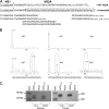
Mass spectrometry analysis of NS1 and NS1′ proteins shows that NS1′ is the product of −1 ribosomal frameshifting. (A) Amino acid sequence of the C-terminal region of NS1 (underlined) and N-terminal region of NS2A with the potential frameshift sequence. Arrows indicate cleavage between the NS1 and NS2A proteins. Frameshift amino acids are enclosed in the dashed box, and the sequences of frameshift peptides detected by mass spectrometry are indicated after trypsin and AspN digestion. (B) Peaks of three of the frameshift peptides detected after AspN digestion of NS1′ (bottom panels) but not of NS1 (top panels). (C) Western blot detection with NS1′ frameshift peptide-specific (FS ab) and 4G4 antibodies. Lysates from Vero and C6/36 cells harvested at 3 and 5 days after WNV infection were separated by 10% PAGE, transferred onto nitrocellulose membranes, and incubated with 4G4 (NS1/NS1′ specific) or FS ab (NS1′ specific).
FIG. 3.
FIG. 3.
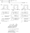
Sequence requirements for −1 ribosomal frameshift in vitro. (A) Diagram of the dual-luciferase reporter system and predicted structures of the WT and mutant sequences used to evaluate the role of WNV, KUNV, and JEV viral sequences in −1 ribosomal frameshifting. Production of an ∼101-kDa product is observed if the inserted sequence induces frameshifting. Locations of different stimulatory elements and mutations are shown as underlined or highlighted sequences. Putative interactions between bases are shown as dashed lines. Frameshifting efficiencies calculated from the intensity of labeled bands in panel B, after normalization for the number of methionine residues in each product, are shown next to the lane numbers in parentheses. (B) SDS-PAGE of in vitro translation products of indicated constructs performed in reticulocyte lysates. The molecular masses of Renilla luciferase (no frameshift, ∼44 kDa) and the frameshift product (∼101 kDa; fusion of Renilla and firefly luciferases) are indicated.
FIG. 4.
FIG. 4.
Sequence requirements for the production of NS1′ and its role in virus infection. (A) Predicted structure(s) of wild-type and mutated heptanucleotide or pseudoknot-forming sequences in KUNV. Altered nucleotides are highlighted, and stimulatory elements are underlined. Predicted interactions between bases in the pseudoknot structure are shown as dashed lines. (B) Western blot detection of NS1 and NS1′ with 4G4 or FS antibodies in lysates from Vero cells infected with WT or mutant KUNV viruses. Lysates from WT-, A30P-, A30A′-, or FSSM-infected cells were separated by PAGE and transferred onto nitrocellulose membranes followed by detection with 4G4 or FS antibodies as in Fig. 2C. (C) Kinetics of replication of WT, A30P, A30A′, and FSSM viruses in BHK and C6/36 cells. Cells were infected at an MOI of 1, and viral accumulation was measured up to 96 h after inoculation using plaque assays of harvested culture fluids. Growth kinetics from a typical experiment are shown. (D) Neuroinvasiveness of mutant viruses in mice. Swiss outbred weanling mice (18 to 19 days old) were inoculated with 1,000 or 10,000 PFU of WT KUNV or mutant viruses. Survival rates were recorded daily up to day 15 after inoculation, when the experiments were terminated. Shown are compiled results from multiple (up to 4) experiments involving WT KUNV (1,000 PFU, 20 mice; 10,000 PFU, 26 mice), KUNV A30P (1,000 PFU, 10 mice; 10,000 PFU, 10 mice), KUNV A30A′ (1,000 PFU, 24 mice; 10,000 PFU, 19 mice), and KUNV FSSM (1,000 PFU, 8 mice; 10,000 PFU, 10 mice).

Similar articles

Cited by

References

    1. Bekaert, M., A. E. Firth, Y. Zhang, V. N. Gladyshev, J. F. Atkins, and P. V. Baranov. 25 September 2009. Recode-2: new design, new search tools, and many more genes. Nucleic Acids Res. [Epub ahead of print.] doi:10.1093/nar/gkp788. - DOI - PMC - PubMed
    1. Blitvich, B. J., J. S. Mackenzie, R. J. Coelen, M. J. Howard, and R. A. Hall. 1995. A novel complex formed between the flavivirus E and NS1 proteins: analysis of its structure and function. Arch. Virol. 140:145-156. - PubMed
    1. Blitvich, B. J., D. Scanlon, B. J. Shiell, J. S. Mackenzie, and R. A. Hall. 1999. Identification and analysis of truncated and elongated species of the flavivirus NS1 protein. Virus Res. 60:67-79. - PubMed
    1. Brierley, I., A. J. Jenner, and S. C. Inglis. 1992. Mutational analysis of the “slippery-sequence” component of a coronavirus ribosomal frameshifting signal. J. Mol. Biol. 227:463-479. - PMC - PubMed
    1. Chen, L. K., C. L. Liao, C. G. Lin, S. C. Lai, C. I. Liu, S. H. Ma, Y. Y. Huang, and Y. L. Lin. 1996. Persistence of Japanese encephalitis virus is associated with abnormal expression of the nonstructural protein NS1 in host cells. Virology 217:220-229. - PubMed

Publication types

MeSH terms

LinkOut - more resources