. 2010 Feb;84(3):1550-62.
doi: 10.1128/JVI.01788-09. Epub 2009 Nov 11.

Attenuation of vesicular stomatitis virus encephalitis through microRNA targeting

Affiliations

Attenuation of vesicular stomatitis virus encephalitis through microRNA targeting

Elizabeth J Kelly et al. J Virol. 2010 Feb.

Abstract

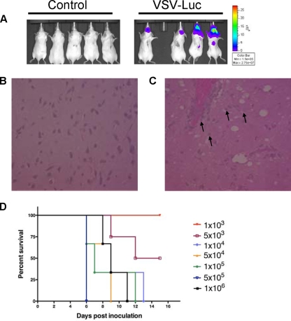
Vesicular stomatitis virus (VSV) has long been regarded as a promising recombinant vaccine platform and oncolytic agent but has not yet been tested in humans because it causes encephalomyelitis in rodents and primates. Recent studies have shown that specific tropisms of several viruses could be eliminated by engineering microRNA target sequences into their genomes, thereby inhibiting spread in tissues expressing cognate microRNAs. We therefore sought to determine whether microRNA targets could be engineered into VSV to ameliorate its neuropathogenicity. Using a panel of recombinant VSVs incorporating microRNA target sequences corresponding to neuron-specific or control microRNAs (in forward and reverse orientations), we tested viral replication kinetics in cell lines treated with microRNA mimics, neurotoxicity after direct intracerebral inoculation in mice, and antitumor efficacy. Compared to picornaviruses and adenoviruses, the engineered VSVs were relatively resistant to microRNA-mediated inhibition, but neurotoxicity could nevertheless be ameliorated significantly using this approach, without compromise to antitumor efficacy. Neurotoxicity was most profoundly reduced in a virus carrying four tandem copies of a neuronal mir125 target sequence inserted in the 3'-untranslated region of the viral polymerase (L) gene.


Figures

FIG. 1.
FIG. 1.
Vesicular stomatitis virus causes lethal neurotoxicity. (A) VSV-Luc localizes to and replicates in the brains of mice. Control Opti-MEM carrier or 1 × 10⁴ VSV-Luc particles were injected intracranially into SCID mice and imaged for luminescence at 7 days postinoculation. (B) H&E-stained section of brain in Opti-MEM-injected control mouse, shown at original magnification of ×400. (C) H&E-stained section of brain of VSV-Luc-injected mouse, shown at original magnification of ×400. Black arrows represent apoptosing neurons. (D) LD50 determination with VSV-Luc administered intracranially. Kaplan-Meier survival curves of dose escalation in SCID mice are shown.
FIG. 2.
FIG. 2.
Analysis of lentiviral vectors incorporating neuron-specific microRNA targets in a panel of cell lines. (A) Schematic diagram of lentiviral vector containing microRNA targets. LTR, long terminal repeat; GA, Gag; SFFV, spleen focus-forming virus (promoter); RRE, Rev response element; cPPT, central polypurine tract. (B) Sequences of inserted target elements. (C) Relative light units (RLU) in control or neuronal cell lines transduced at an MOI of 1.0 with lentiviral vectors containing miR-142-3pT, miR-124T, miR-125T, miR-128T, or miR-134T or in the presence of 200 nM sequence-complementary miRNA mimic. (D) miRNA expression profiling determined by TaqMan expression analysis. Fold increases indicate enrichment over miRNA expression in HeLa cells (per 5 ng small RNA). Gray asterisks, P < 0.05; black asterisks, P < 0.01.
FIG. 3.
FIG. 3.
Construction and characterization of recombinant VSVs. (A) Schematic diagram of microRNA-targeted VSV. A novel NotI restriction site was created in the 3′UTR of the M, Luc, or L gene, and miRTs were cloned into this site. (B) One-step growth curves for recombinant viruses on BHK cells.
FIG. 4.
FIG. 4.
VSVs encoding neuronal targets are largely unresponsive to sequence-complementary miRNA mimics. (A) Cell viability, as determined by MTT assay, at 24 h post-viral infection at an MOI of 0.5 with recombinant miRNA-targeted VSVs pretreated for 4 h with 200 nM miRNA mimics in A549 cells. (B) Corresponding luciferase activities of miRT VSVs in the presence of the miRNA mimics from panel A. (C) Viral titers collected at 24 h postinfection from supernatants of cells infected with miRT VSVs in the presence of miRNA mimics (representative of four repeat experiments). #, not determined; *, P = 0.0012.
FIG. 5.
FIG. 5.
Viral replication of miRT VSVs is restricted in primary brain cells. (A) Cell viability, as determined by MTT assay, at 24 h post-viral infection at an MOI of 0.5 with recombinant miRNA-targeted VSVs in primary astrocytes. (B) Viral titers collected at 24 h postinfection from supernatants of cells infected with miRT VSVs in primary astrocytes (representative of three repeat experiments). (C) miRNA abundance in primary astrocytes, as determined by TaqMan miRNA expression analysis. Fold increases in miR-124, miR-125, or miR-206 over corresponding miRNAs in HeLa cells (per 5 ng small RNA) are shown. *, P < 0.001.
FIG. 6.
FIG. 6.
VSVs encoding neuron-specific miR-125T have reduced neurotoxicity. Mice were inoculated intracranially with 1 × 10⁴ rVSV particles and were imaged at the indicated times, using an IVIS200 system (Xenogen Corp., Alameda, CA), and monitored for symptoms of neurotoxicity. (A) VSV-Luc at 7 days postinfection (p.i.). (B) VSV 125r M at 7 days p.i. (C) VSV 125r L at 7 days p.i. (D) VSV 206r L at 7 days p.i. (E) Kaplan-Meier survival graphs for mice inoculated intracranially with 1 × 10⁴ rVSV particles. (F) Quantification of the amount of bioluminescence output per brain, plotted over time, as an indication of viral gene expression for mice inoculated with rVSV (values are averages for 10 animals per group ± standard deviations). (G) A mouse administered 1 × 10⁴ VSV 125 L particles (negative until imaged on day 28) developed encephalitis, as seen at the labeled times postinfection. *, P = 0.0025.
FIG. 7.
FIG. 7.
MicroRNA-targeted VSVs have equivalent antitumor activities in CT-26 model. (A) CT-26 cells (5 × 10⁶) were injected subcutaneously into the right flank of BALB/c mice, and mice were administered one intratumoral dose of virus (1 × 10⁹) on day 0 and one intravenous dose (1 × 10⁹) on day 3 and monitored for tumor progression. (B) Kaplan-Meier survival curves for the mice in panel A. Images show CT-26 tumor-bearing mice treated with rVSV on day 2, and graphs show the quantification of bioluminescence output per flank, plotted over time, as an indication of viral gene expression for mice treated with VSV-Luc (C), VSV 125r M (D), VSV 125r L (E), and VSV 206r L (F).

References

    1. Altomonte, J., R. Braren, S. Schulz, S. Marozin, E. J. Rummeny, R. M. Schmid, and O. Ebert. 2008. Synergistic antitumor effects of transarterial viroembolization for multifocal hepatocellular carcinoma in rats. Hepatology 48:1864-1873. - PubMed
    1. Balachandran, S., and G. N. Barber. 2004. Defective translational control facilitates vesicular stomatitis virus oncolysis. Cancer Cell 5:51-65. - PubMed
    1. Ball, L. A., C. R. Pringle, B. Flanagan, V. P. Perepelitsa, and G. W. Wertz. 1999. Phenotypic consequences of rearranging the P, M, and G genes of vesicular stomatitis virus. J. Virol. 73:4705-4712. - PMC - PubMed
    1. Barber, G. N. 2005. VSV-tumor selective replication and protein translation. Oncogene 24:7710-7719. - PubMed
    1. Barnes, D., M. Kunitomi, M. Vignuzzi, K. Saksela, and R. Andino. 2008. Harnessing endogenous miRNAs to control virus tissue tropism as a strategy for developing attenuated virus vaccines. Cell Host Microbe 4:239-248. - PMC - PubMed

LinkOut - more resources