. 2010 Feb;84(3):1334-47.
doi: 10.1128/JVI.01301-09. Epub 2009 Nov 11.

Wide-scale use of Notch signaling factor CSL/RBP-Jkappa in RTA-mediated activation of Kaposi's sarcoma-associated herpesvirus lytic genes

Affiliations

Wide-scale use of Notch signaling factor CSL/RBP-Jkappa in RTA-mediated activation of Kaposi's sarcoma-associated herpesvirus lytic genes

Linda M Persson et al. J Virol. 2010 Feb.

Abstract

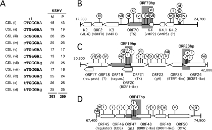
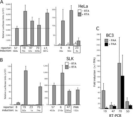
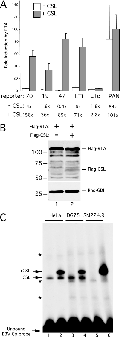
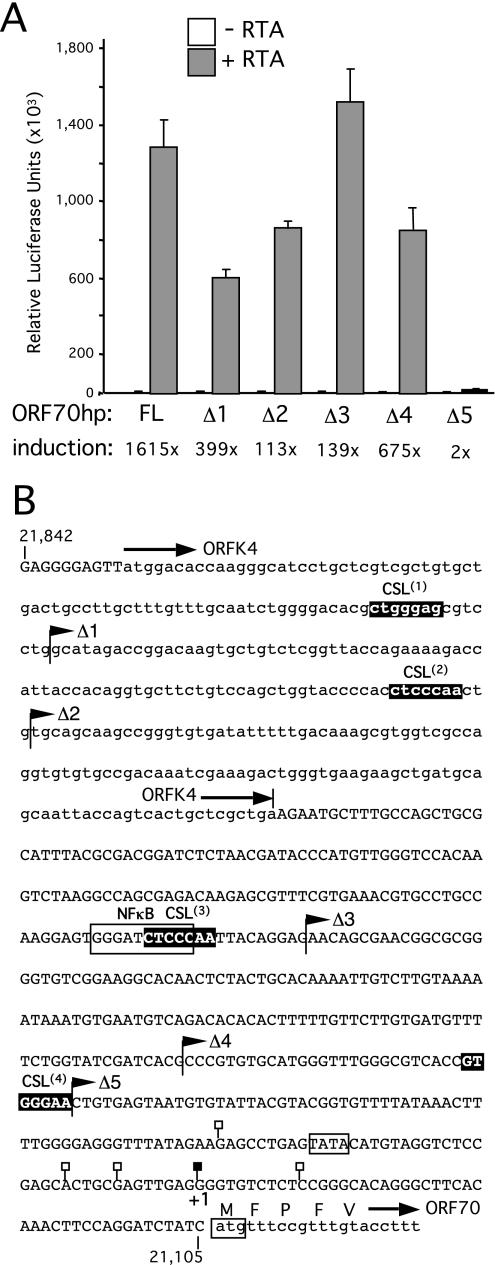
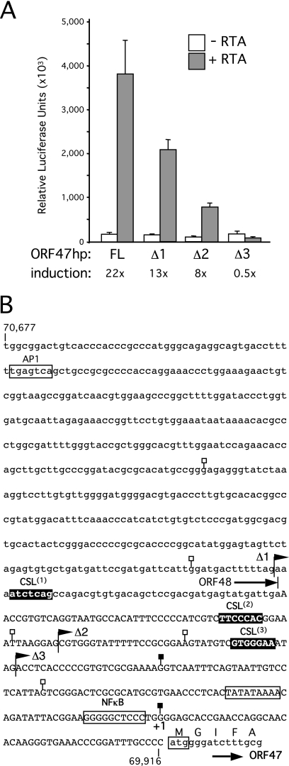
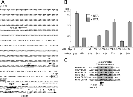
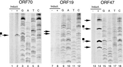
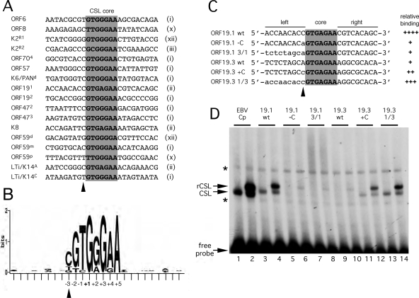
For Kaposi's sarcoma-associated herpesvirus (KSHV; also called human herpesvirus 8 [HHV8]), the switch from latency to active lytic replication requires RTA, the product of open reading frame 50 (ORF50). RTA activates transcription from nearly 40 early and delayed-early viral promoters, mainly through interactions with cellular DNA binding proteins, such as CSL/RBP-Jkappa, Oct-1, C/EBPalpha, and c-Jun. Reliance on cellular coregulators may allow KSHV to adjust its lytic program to suit different cellular contexts or interpret signals from the outside. CSL is a key component of the Notch signaling pathway and is targeted by several viruses. A search with known CSL binding sequences from cellular genes found at least 260 matches in the KSHV genome, many from regions containing known or suspected lytic promoters. Analysis of clustered sites located immediately upstream of ORF70 (thymidylate synthase), ORF19 (tegument protein), and ORF47 (glycoprotein L) uncovered RTA-responsive promoters that were validated using mRNAs isolated from KSHV-infected cells undergoing lytic reactivation. Notably, ORF19 behaves as a true late gene, indicating that RTA regulates all three phases of the lytic program. For each new promoter, the response to RTA was dependent on CSL, and 5 of the 10 candidate sites were shown to bind CSL in vitro. Analysis of individual sites highlighted the importance of a cytosine residue flanking the core CSL binding sequence. These findings broaden the role for CSL in coordinating the KSHV lytic gene expression program and help to define a signature motif for functional CSL sites within the viral genome.


Figures

FIG. 1.
FIG. 1.
The KSHV genome contains many matches to known CSL binding sites. (A) Published studies of mammalian Notch-regulated genes describe 11 variants (i through xi) of the canonical CSL recognition sequence. The numbers of matches to each variant present in the M-type (49) (NCBI accession no. NC_003409) and P-type (17) (NCBI accession no. NC_009333) KSHV genome sequences are indicated on the right. (B) Schematic showing 7.5-kbp region (positions 17,200 to 24,700 in the M-type reference sequence) between ORF K2 and the intergenic region containing the lytic origin of replication. The positions and type of potential CSL sites are indicated by lollipops. The region immediately upstream of ORF70 (ORF70hp; shaded), containing four candidate sites, was selected for functional analysis. (C) Map of the 12-kbp region (positions 30,800 to 42,800) between ORF17 and ORF24, containing ORF19hp and ORF23hp. (D) Region (7.5 kbp) between ORF45 and ORF50 (positions 67,400 to 74,900), including ORF47hp.
FIG. 2.
FIG. 2.
Identification of novel RTA-responsive promoters in the intergenic regions upstream of ORF70, ORF19, and ORF47. Reporter constructs were generated by subcloning the following fragments into a promoterless luciferase reporter: ORF6 (M-type reference sequence nucleotides [nt] 2,472 to 3,209), ORF8 (nt 7,959 to 8,698), ORF70hp (nt 21,105 to 21,842), ORF23 (nt 40,517 to 41,287), ORF19hp (nt 34,844 to 35,583), ORF47 (nt 69,916 to 70,677), and ORF57 (nt 81,331 to 82,086). Activity was assayed by transient transfection of HeLa (A) and SLK (B) cells by cotransfection with a full-length RTA (pCGFlag-RTAFL) expression plasmid or the empty equivalent. Each assay was performed in triplicate, and relative luciferase activity was measured after 24 h. For each combination, the mean and standard deviation are shown, together with the calculated difference between the presence and absence of RTA (fold induction). Two previously characterized RTA-responsive reporters, LTi-luc (nucleotides 127,610 to 127,807) and PAN-luc (nucleotides 28,455 to 28,681), were included as positive controls (42). ORF57-luc showed similar levels of induction in the two cell types, and its activity is used as a point of comparison (dotted line). (C) Latently infected BC3 cells were induced with TPA in the presence or absence of PAA. After 72 h, total RNA was isolated and analyzed by RT-PCR, using primers specific to ORF19, ORF47, ORF70, and ORF50/RTA. Values represent the mean and standard deviations obtained from assaying three independent cultures. Differences in RNA handling and cDNA synthesis were normalized using 18S rRNA.
FIG. 3.
FIG. 3.
CSL is required for response to RTA. (A) Analysis of induction by RTA in CSL-null SM224.9 cells, a derivative of the DG75 cell line carrying a somatic knockout of the CSL gene (41). Plasmid DNA was introduced into 0.5 × 10⁷ SM224.9 cells by electroporation, and luciferase activity was determined after 24 h. Each reporter plasmid, ORF70hp-luc, ORF19hp-luc, ORF47hp-luc, LTi-luc, LTc-luc, and PAN-luc, was cotransfected with combinations of empty vector, RTA vector, or CSL vector. Fold induction by RTA was calculated for each reporter, in either the presence (+) or absence (−) of cotransfected CSL. The mean and standard deviation of three separate assays are shown. (B) Immunoblot of whole-cell extracts prepared from SM224.9 cells transfected with plasmids encoding Flag-RTA (lane 1) or Flag-RTA and Flag-CSL (lane 2). Extracts were separated in an SDS-10% PAGE gel and probed with anti-FLAG or anti-Rho-GDI antibodies. (C) Gel shift assay using whole-cell extracts prepared from HeLa (lanes 1 and 2), DG75 (lanes 3 and 4), and SM224.9 (lanes 5 and 6) cells that were either mock transfected (lanes 1, 3, and 5) or transfected with a plasmid encoding epitope-tagged CSL (lanes 2, 4, and 6). Each extract was mixed with a 32P-labeled probe containing the canonical CSL binding site from the EBV Cp promoter, incubated for 30 min at 30°C, and resolved in a 4% native PAGE gel. Positions of the unbound (free) probe and shifted complexes corresponding to endogenous (CSL) and transfected (rCSL) CSL are indicated. Nonspecific complexes are indicated with asterisks.
FIG. 4.
FIG. 4.
Analysis of ORF70-K4 intergenic region. (A) Full-length (FL) and truncated (Δ1, Δ2, Δ3, Δ4, and Δ5) versions of ORF70hp-luc were tested for a response to RTA in HeLa cells by transient transfection. Assays were performed in triplicate and the luciferase values expressed as the mean and standard deviation. For each reporter, fold induction was calculated from luciferase activity measured in the presence and absence of the RTA expression plasmid. (B) Annotated sequence of the ORF70hp fragment (nucleotides 21,105 to 21,842 in the M-type reference sequence). Overlap with ORFK4 (encoding vMIP2) is shown in lowercase, as are the first few codons of ORF70, encoding thymidylate synthase. Also shown are the end points of the truncations used in panel A (filled flags), the four candidate CSL sites, ORF70.1, ORF70.2, ORF70.3, and ORF70.4 (filled boxes), and TSSs mapped by 5′-RACE (filled or open lollipops). The principal TSS (filled lollipop) is labeled +1 and corresponds to position 21,150. A putative TATA box and consensus NF-κB site are indicated with an open box.
FIG. 5.
FIG. 5.
Truncation analysis of the ORF47 promoter region. (A) Analysis of full-length (FL) and truncated (Δ1, Δ2, and Δ3) versions of ORF47hp-luc in HeLa cells. (B) Sequence of the ORF47hp fragment (nucleotides 69,916 to 70,677). The region of overlap with ORF48 (EBV BRRF2 homolog) is shown in lowercase, as are the first five codons of ORF47. Also shown are the end points of truncations used in panel A (filled flags), the three candidate CSL sites, ORF47.1, ORF47.2, and ORF47.3 (filled boxes), and TSSs mapped by 5′-RACE (filled or open lollipops). The preferred TSS (filled lollipop) is labeled +1 and corresponds to position 69,963 in the KSHV prototype genome. Consensus AP1 and NF-κB binding sequences and a putative TATA box are also indicated.
FIG. 6.
FIG. 6.
Analysis of ORF19-ORF21 intergenic region. (A) Sequence of the ORF19hp fragment (nucleotides 34,844 to 35,583). The 5′ end of ORF21 (thymidine kinase), encoded on the opposite strand from ORF19, is shown in lowercase, as are the first five codons of ORF19. Also shown are the end points of truncations used in panel B (filled flags) and the three candidate CSL sites, ORF19.1, ORF19.2, and ORF19.3 (filled boxes). Primer extension product end points were mapped to a region around the second CSL site (dotted line). A consensus AP1 site is indicated with an open box. (B) Full-length (FL) ORF19hp-luc and a series of truncations (Δ1 and Δ2) and targeted mutations were tested for a response to RTA in HeLa cells by transient transfection. Assays were performed in triplicate and luciferase values expressed as the mean and standard deviation. For each reporter, fold induction was calculated from luciferase activities measured in the presence and absence of the RTA expression plasmid. (C) Alignment of the ORF19 TA-rich element with similar sequences found in the Epstein-Barr virus (EBV) BcLF1 (51), human cytomegalovirus (HCMV) UL94 (68), HCMV UL75 (43), KSHV K8.1 (56), and KSHV ORF17.5 (5) true late promoters. Shading indicates the 12-bp core element defined by studies of the K8.1 promoter, with conserved nucleotides shown in bold.
FIG. 7.
FIG. 7.
Mapping the 5′ ends of RTA-inducible transcripts. Poly(A)+ RNAs from induced (+) and uninduced (−) rKSHV.219 Vero cells were annealed to 32P-labeled antisense oligonucleotides specific to ORF70 (lanes 1 and 2), ORF19 (lanes 7 and 8), or ORF47 (lanes 13 and 14). Primers were extended using reverse transcriptase, and the denatured products were resolved in an 8% sequencing gel. The sizes of the extension products were determined using radiolabeled molecular weight markers and a sequencing ladder generated with the same labeled primer and a subcloned fragment of KSHV genomic DNA as the sequencing template. Extension products that coincide with end points detected by 5′-RACE are indicated with lollipops.
FIG. 8.
FIG. 8.
In vitro binding analysis of candidate CSL binding sites. Gel mobility shift assays were performed using 32P-labeled probes containing candidate CSL binding sites from the ORF70 (A), ORF19 (B), and ORF47 (C) promoters. Individual sites are numbered in accordance with Fig. 4, 5, and 6. Characterized binding sites from the EBV Cp promoter (panel A, lanes 1 and 2) and the ORF57 promoter (panel B, lanes 1 and 2) were included as positive controls. All probes were identical in length and were mixed with HeLa whole-cell extract from mock-transfected cells (odd-numbered lanes) or cells expressing tagged CSL (even-numbered lanes). Binding reaction mixtures were incubated at 30°C for 30 min prior to being loaded on a 4% native PAGE gel. Positions of the unbound probe and complexes corresponding to endogenous (CSL) or transfected CSL (rCSL) are indicated. Nonspecific complexes are marked with asterisks.
FIG. 9.
FIG. 9.
Defining a signature for CSL binding sites in the KSHV genome. (A) Alignment of 17 confirmed CSL binding sites from the KSHV genome. The core sequence is shaded, and the core motif type, as defined in Fig. 1A, is noted on the right. Labeling of individual sites from K2/vIL6, K6/PAN, ORF59, and LTi/K14 follows published naming schemes (4, 33, 34, 36). (B) A positional nucleotide frequency pattern (sequence logo) for the sites listed in panel A was generated using WebLogo (9). Only positions +1, +3, and +5 are invariant. This visual representation emphasizes the strong preference for a cytosine at position −3 and the striking absence of sequence conservation beyond the eight central positions (−3 to +5). (C) Sequences of probes used to test the contributions of flanking residues to the differential binding of CSL to ORF19 sites 1 and 3. The wild-type sequence is shown in uppercase, and the altered sequence is shown in lowercase. (D) Gel mobility shift analysis of the probes shown in panel C. Each was labeled to an identical specific activity and then incubated with HeLa whole-cell extract from mock-transfected cells (odd-numbered lanes) or cells expressing tagged CSL (even-numbered lanes). The EBV Cp site (lanes 1 and 2) was included as a reference. Positions of the unbound probe and complexes corresponding to endogenous (CSL) or transfected (rCSL) CSL are indicated. Nonspecific complexes are marked with asterisks. The ability of each probe to form a stable complex with CSL is summarized in panel C.

Similar articles

Cited by

References

    1. Borggrefe, T., and F. Oswald. 2009. The Notch signaling pathway: transcriptional regulation at Notch target genes. Cell Mol. Life Sci. 66:1631-1646. - PMC - PubMed
    1. Bray, S. J. 2006. Notch signalling: a simple pathway becomes complex. Nat. Rev. Mol. Cell Biol. 7:678-689. - PubMed
    1. Carroll, K. D., F. Khadim, S. Spadavecchia, D. Palmeri, and D. M. Lukac. 2007. Direct interactions of KSHV/HHV-8 ORF50/Rta protein with the cellular protein octamer-1 and DNA are critical for specifying transactivation of a delayed-early promoter and stimulating viral reactivation. J. Virol. 81:8451-8467. - PMC - PubMed
    1. Chang, H., D. P. Dittmer, S. Y. Chul, Y. Hong, and J. U. Jung. 2005. Role of Notch signal transduction in Kaposi's sarcoma-associated herpesvirus gene expression. J. Virol. 79:14371-14382. - PMC - PubMed
    1. Chang, J., and D. Ganem. 2000. On the control of late gene expression in Kaposi's sarcoma-associated herpesvirus (human herpesvirus-8). J. Gen. Virol. 81:2039-2047. - PubMed

Publication types

MeSH terms

Substances

LinkOut - more resources