Chimeric feline coronaviruses that encode type II spike protein on type I genetic background display accelerated viral growth and altered receptor usage
- PMID: 19906918
- PMCID: PMC2812337
- DOI: 10.1128/JVI.01568-09
Chimeric feline coronaviruses that encode type II spike protein on type I genetic background display accelerated viral growth and altered receptor usage
Abstract
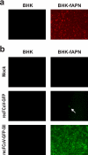
Persistent infection of domestic cats with feline coronaviruses (FCoVs) can lead to a highly lethal, immunopathological disease termed feline infectious peritonitis (FIP). Interestingly, there are two serotypes, type I and type II FCoVs, that can cause both persistent infection and FIP, even though their main determinant of host cell tropism, the spike (S) protein, is of different phylogeny and displays limited sequence identity. In cell culture, however, there are apparent differences. Type II FCoVs can be propagated to high titers by employing feline aminopeptidase N (fAPN) as a cellular receptor, whereas the propagation of type I FCoVs is usually difficult, and the involvement of fAPN as a receptor is controversial. In this study we have analyzed the phenotypes of recombinant FCoVs that are based on the genetic background of type I FCoV strain Black but encode the type II FCoV strain 79-1146 S protein. Our data demonstrate that recombinant FCoVs expressing a type II FCoV S protein acquire the ability to efficiently use fAPN for host cell entry and corroborate the notion that type I FCoVs use another main host cell receptor. We also observed that recombinant FCoVs display a large-plaque phenotype and, unexpectedly, accelerated growth kinetics indistinguishable from that of type II FCoV strain 79-1146. Thus, the main phenotypic differences for type I and type II FCoVs in cell culture, namely, the growth kinetics and the efficient usage of fAPN as a cellular receptor, can be attributed solely to the FCoV S protein.
Figures


Similar articles
-
Feline Coronaviruses: Pathogenesis of Feline Infectious Peritonitis.Adv Virus Res. 2016;96:193-218. doi: 10.1016/bs.aivir.2016.08.002. Epub 2016 Aug 31. Adv Virus Res. 2016. PMID: 27712624 Free PMC article. Review.
-
A Tale of Two Viruses: The Distinct Spike Glycoproteins of Feline Coronaviruses.Viruses. 2020 Jan 10;12(1):83. doi: 10.3390/v12010083. Viruses. 2020. PMID: 31936749 Free PMC article. Review.
-
Tackling feline infectious peritonitis via reverse genetics.Bioengineered. 2014;5(6):396-400. doi: 10.4161/bioe.32133. Epub 2014 Oct 30. Bioengineered. 2014. PMID: 25482087 Free PMC article.
-
Establishment of a Virulent Full-Length cDNA Clone for Type I Feline Coronavirus Strain C3663.J Virol. 2019 Oct 15;93(21):e01208-19. doi: 10.1128/JVI.01208-19. Print 2019 Nov 1. J Virol. 2019. PMID: 31375588 Free PMC article.
-
Limitations of using feline coronavirus spike protein gene mutations to diagnose feline infectious peritonitis.Vet Res. 2017 Oct 5;48(1):60. doi: 10.1186/s13567-017-0467-9. Vet Res. 2017. PMID: 28982390 Free PMC article.
Cited by
-
Feline Coronaviruses: Pathogenesis of Feline Infectious Peritonitis.Adv Virus Res. 2016;96:193-218. doi: 10.1016/bs.aivir.2016.08.002. Epub 2016 Aug 31. Adv Virus Res. 2016. PMID: 27712624 Free PMC article. Review.
-
Establishment and Cross-Protection Efficacy of a Recombinant Avian Gammacoronavirus Infectious Bronchitis Virus Harboring a Chimeric S1 Subunit.Front Microbiol. 2022 Jul 22;13:897560. doi: 10.3389/fmicb.2022.897560. eCollection 2022. Front Microbiol. 2022. PMID: 35935229 Free PMC article.
-
A Tale of Two Viruses: The Distinct Spike Glycoproteins of Feline Coronaviruses.Viruses. 2020 Jan 10;12(1):83. doi: 10.3390/v12010083. Viruses. 2020. PMID: 31936749 Free PMC article. Review.
-
Feline coronavirus: Insights into viral pathogenesis based on the spike protein structure and function.Virology. 2018 Apr;517:108-121. doi: 10.1016/j.virol.2017.12.027. Epub 2018 Jan 10. Virology. 2018. PMID: 29329682 Free PMC article.
-
Emergence of pathogenic coronaviruses in cats by homologous recombination between feline and canine coronaviruses.PLoS One. 2014 Sep 2;9(9):e106534. doi: 10.1371/journal.pone.0106534. eCollection 2014. PLoS One. 2014. PMID: 25180686 Free PMC article.
References
Publication types
MeSH terms
Substances
LinkOut - more resources
Full Text Sources
Miscellaneous

