. 2010 Jan;84(2):773-87.
doi: 10.1128/JVI.01190-09. Epub 2009 Nov 11.

Identification of GBF1 as a cellular factor required for hepatitis C virus RNA replication

Affiliations

Identification of GBF1 as a cellular factor required for hepatitis C virus RNA replication

Lucie Goueslain et al. J Virol. 2010 Jan.

Abstract

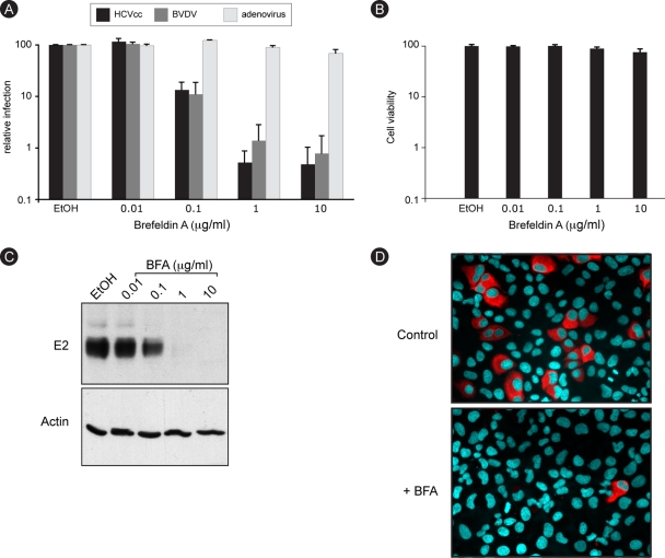
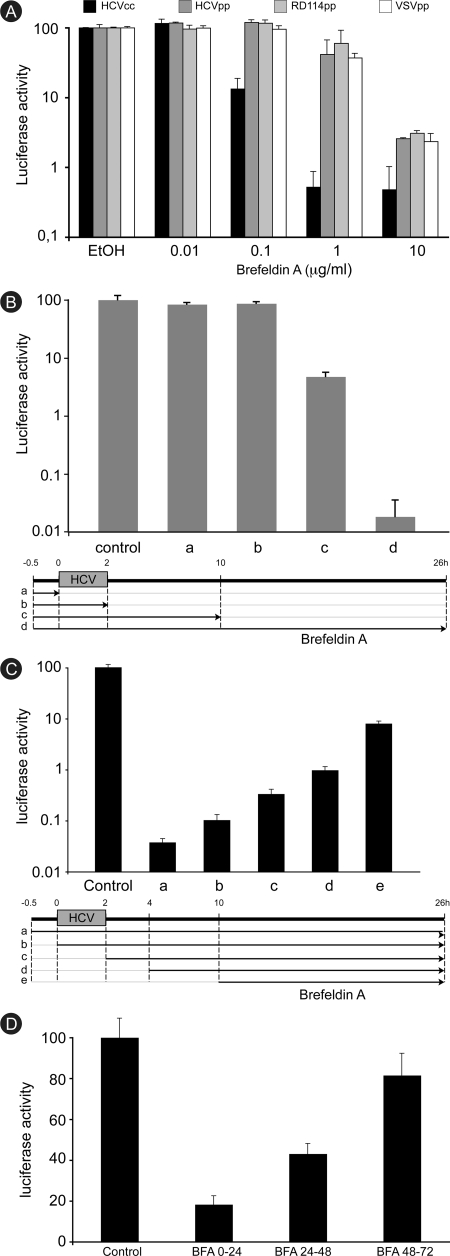
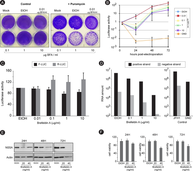
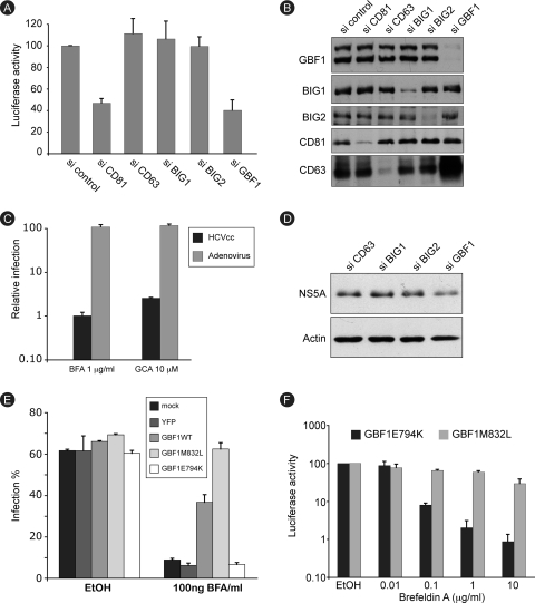
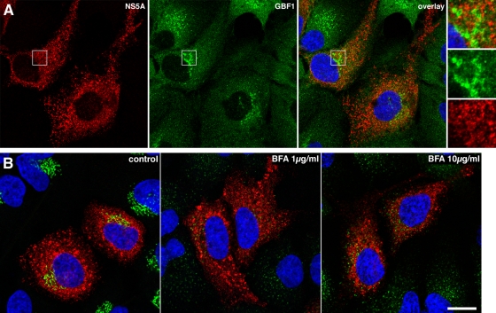
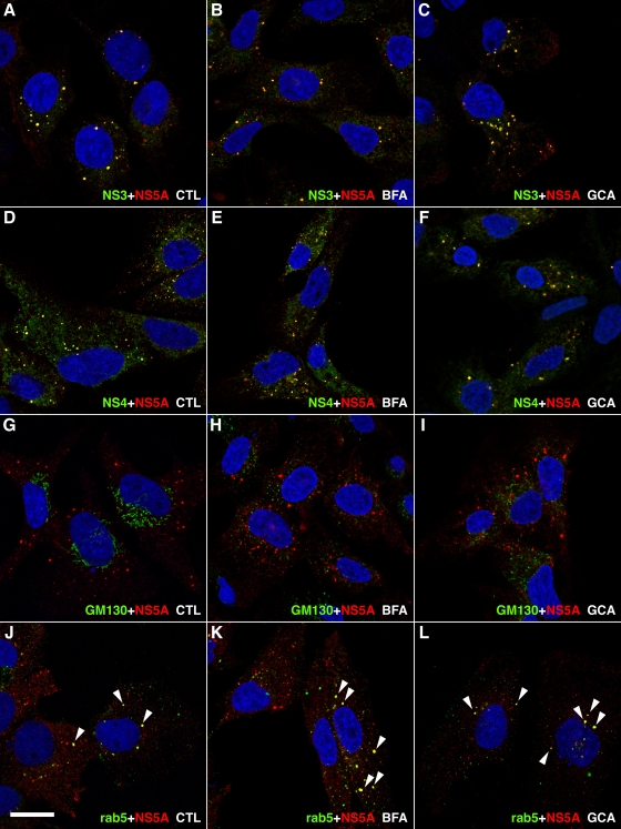
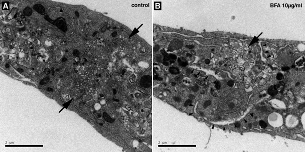
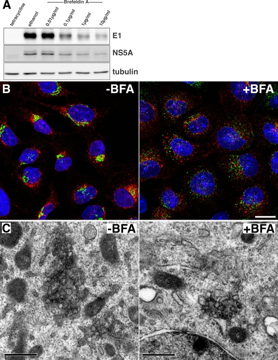
In infected cells, hepatitis C virus (HCV) induces the formation of membrane alterations referred to as membranous webs, which are sites of RNA replication. In addition, HCV RNA replication also occurs in smaller membrane structures that are associated with the endoplasmic reticulum. However, cellular mechanisms involved in the formation of HCV replication complexes remain largely unknown. Here, we used brefeldin A (BFA) to investigate cellular mechanisms involved in HCV infection. BFA acts on cell membranes by interfering with the activation of several members of the family of ADP-ribosylation factors (ARF), which can lead to a wide range of inhibitory actions on membrane-associated mechanisms of the secretory and endocytic pathways. Our data show that HCV RNA replication is highly sensitive to BFA. Individual knockdown of the cellular targets of BFA using RNA interference and the use of a specific pharmacological inhibitor identified GBF1, a guanine nucleotide exchange factor for small GTPases of the ARF family, as a host factor critically involved in HCV replication. Furthermore, overexpression of a BFA-resistant GBF1 mutant rescued HCV replication in BFA-treated cells, indicating that GBF1 is the BFA-sensitive factor required for HCV replication. Finally, immunofluorescence and electron microscopy analyses indicated that BFA does not block the formation of membranous web-like structures induced by expression of HCV proteins in a nonreplicative context, suggesting that GBF1 is probably involved not in the formation of HCV replication complexes but, rather, in their activity. Altogether, our results highlight a functional connection between the early secretory pathway and HCV RNA replication.


Figures

FIG. 1.
FIG. 1.
HCV infection is sensitive to BFA. (A) Huh-7 cells were infected with HCV or GFP-expressing adenovirus, and MDBK cells were infected with BVDV in the presence of 0.2% ethanol (EtOH) or increasing concentrations of BFA. BFA was present for 8 h for Huh-7 cells (HCVcc and adenovirus) or throughout the experiment for MDBK cells (BVDV). At 24 h postinfection, cells were harvested for luciferase assays (HCVcc) or fluorescence-activated cell sorter analysis (BVDV and adenovirus). The luciferase activity or number of infected cells for ethanol-treated cells is expressed as 100%. Error bars indicate standard errors of the means for 4 (HCV), 6 (BVDV), or 3 (adenovirus) experiments. (B) Huh-7 cells were incubated for 8 h in the presence of 0.2% EtOH or increasing concentrations of BFA and then cultured for 18 h without drug. Viability was assessed using an MTS assay. The absorbance of the ethanol-treated sample is expressed as 100%. (C) Huh-7 cells were infected with HCVcc as for panel A. At 30 h postinfection, E2 and actin expression levels were analyzed by immunoblotting. (D) Huh-7 cells were infected for 2 h with HCVcc in the presence of 0.2% EtOH or 1 μg/ml BFA. BFA was removed at 6 h postinfection. Cells were fixed at 30 h postinfection and processed for immunofluorescence detection of E2.
FIG. 2.
FIG. 2.
Brefeldin A inhibits a postentry step of HCV infection. (A) Huh-7 cells were infected with HCVcc, HCVpp, RD114pp, or VSVpp in the presence of 0.2% ethanol (EtOH) or increasing concentrations of BFA. BFA was removed at 6 h postinfection. Luciferase activity was measured at 24 h or 48 h postinfection for HCVcc and pseudoparticles, respectively. The luciferase activity from ethanol-treated cells is expressed as 100%. Error bars indicate standard deviations. (B) Huh-7 cells were pretreated for 30 min with 50 ng/ml BFA and infected for 2 h. BFA was removed either before infection (a), just after infection (b), or at 8 h postinfection (c) or was present throughout the experiment (d). Luciferase activity was measured at 24 h postinfection. The luciferase activity from untreated cells (control) is expressed as 100%. (C) BFA was added to HCVcc-infected cells either 30 min before infection (a), at the beginning of the infection (b), just after infection (c), 2 h postinfection (d), or 8 h postinfection (e). Luciferase activity was measured at 24 h postinfection. The luciferase activity from untreated cells (control) is expressed as 100%. (D) HCVcc-infected cells were treated with 50 ng/ml BFA from 0 to 24 h, from 24 to 48 h, or from 48 to 72 h. Luciferase activity was measured at 72 h postinfection for all the samples. The luciferase activity from untreated cells (control) is expressed as 100%.
FIG. 3.
FIG. 3.
Brefeldin A inhibits RNA replication. (A) Huh-7 cells were electroporated with a recombinant HCV genome containing a deletion in E1E2 and expressing a puromycin acetyltransferase selection marker. Cells were cultured in the presence of increasing concentrations of BFA for 8 h and then in the presence or the absence of 1 μg/ml puromycin for 6 days. Cells were stained with crystal violet. (B) Huh-7 cells were electroporated with a recombinant HCV genome containing a deletion in E1E2 and expressing Renilla luciferase and were cultured in the presence of BFA for 8 h and then in the absence of the drug. For comparison, Huh-7 cells were electroporated with a nonreplicative (GND) HCV genome and cultured in the absence of BFA. Samples were harvested for luciferase assay at 4, 24, 48, and 72 h postelectroporation. The luciferase activity from ethanol-treated cells at 4 h postelectroporation is expressed as 1. Error bars indicate standard errors of the means for 3 experiments. (C) Huh-7 cells were electroporated with in vitro-transcribed and capped RNA constructs expressing firefly luciferase (F-Luc) under cap control and Renilla luciferase (R-Luc) under HCV IRES control. Cells were cultured in the presence of increasing concentrations of BFA and harvested for dual-luciferase assay at 8 h postelectroporation. (D) Huh-7 cells were infected with HCVcc, treated with increasing concentrations of BFA for 8 h, and harvested at 24 h postinfection for quantitative RT-PCR quantification of HCV plus and minus strands. To confirm the specificity of negative-strand amplification, cells electroporated with in vitro-transcribed HCV replicative (JFH1) or nonreplicative (GND) genomes were processed in parallel (control). (E and F) Huh-7 cells harboring a subgenomic replicon were cultured in the presence of the indicated concentrations of BFA. Samples were harvested after 24, 48, or 72 h of treatment for immunoblot detection of NS5A and actin (E) or for analysis of cell viability (F).
FIG. 4.
FIG. 4.
GBF1 is the brefeldin A-sensitive factor required for HCV replication. (A) Huh-7 cells were transfected with the indicated siRNAs and infected with HCV. Luciferase activity was measured at 24 h postinfection. The luciferase activity from control siRNA-transfected cells (si control) is expressed as 100%. Error bars indicate standard errors of the means for 6 experiments. (B) siRNA-mediated depletion of target proteins was verified by immunoblot analysis. (C) Huh-7 cells were infected with HCV-RLuc or GFP-expressing adenovirus in the presence of 1 μg/ml BFA, 0.02% ethanol (BFA stock solvent), 10 μM golgicide A (GCA), or 0.02% dimethyl sulfoxide (DMSO) (golgicide A stock solvent). Both drugs were present for 8 h. At 24 h postinfection, cells were harvested for luciferase assay (HCVcc) or fluorescence-activated cell sorter analysis (adenovirus). The luciferase activity and number of adenovirus-infected cells from ethanol- or DMSO-treated samples are expressed as 100%. (D) Huh-7 cells harboring a subgenomic replicon were transfected with the indicated siRNAs. Cells were lysed, and cell lysates were analyzed by immunoblotting with antibodies to NS5A and actin. (E) Huh-7 cells were transfected with expression plasmids for GBF1, BFA-resistant mutant GBF1-M832L, GBF1 inactive mutant E794K, or YFP. Transfected cells were infected with HCVcc and cultured in the presence or absence of BFA. Cells were fixed at 24 h postinfection and processed for immunofluorescence detection of E1. Results are presented as percentage of infected cells. (F) Huh-7 cells were transfected with expression plasmids for GBF1-M832L or GBF1-E794K, infected with HCV-RLuc in the presence of 0.2% ethanol or the indicated concentration of BFA, and cultured in the presence of BFA for 8 h and then in the absence of the drug. Luciferase activity was measured at 24 h postinfection. The luciferase activity from ethanol-treated cells is expressed as 100%.
FIG. 5.
FIG. 5.
Immunofluorescence analysis of GBF1 localization and BFA effect in HCV-infected cells. (A) HCV-infected cells were fixed and processed for double-label immunofluorescence for NS5A (red) and GBF1 (green) at 24 h postinfection. Representative confocal images are shown together with the merge image and an enlargement of the indicated area. (B) HCV-infected cells were treated for 1 h with the indicated concentration of BFA, fixed, and processed for double-label immunofluorescence for NS5A (red) and the Golgi marker GM130 (green). Representative confocal images are shown.
FIG. 6.
FIG. 6.
Immunofluorescence analysis of GBF1 inhibition in subgenomic replicon-harboring cells. Huh-7 cells harboring a subgenomic replicon were treated for 8 h in the presence of ethanol 0.2% (A, D, G, and J), 1 μg/ml BFA (B, E, H, and K), or 10 μM golgicide A (C, F, I, and L) and processed for double-label immunofluorescence using anti-NS5A antibody together with antibodies to NS3 (A to C), to NS4A-B (D to F), to cis-Golgi marker GM130 (G to I), or to Rab5 (J to L). Representative confocal images are shown, with NS5A in red and other markers in green. Colocalization of both markers in dot-like structures appears in yellow and is marked by white arrowheads in panels J to L. Bar, 20 μm.
FIG. 7.
FIG. 7.
Membrane alterations in BFA-treated cells visualized by electron microscopy. HCVcc-infected Huh-7 cells were incubated for 8 h in the presence of 0.2% ethanol (A) or 10 μg/ml BFA (B) and processed for electron microscopy. Membranous webs are indicated by arrows. Arrowheads indicate examples of swollen ER cisternae in BFA-treated cells. Bars, 2 μm.
FIG. 8.
FIG. 8.
Effect of brefeldin A on membranous web-like structure formation in UHCV-11 cells. (A) UHCV-11 cells were induced by tetracycline withdrawal and incubated for 8 h in the presence of different concentrations of BFA or ethanol (0.2%). Control cells were incubated for 8 h without induction (tetracycline). HCV protein expression was assessed by immunoblot analysis of E1, NS5A, and tubulin. (B) Cells induced for 8 h in the presence or absence of BFA (1 μg/ml) were fixed and processed for immunofluorescence detection of NS5A (red) and the Golgi marker GM130 (green) Nuclei were stained with DAPI (blue). Bar, 20 μm. (C) Cells induced for 8 h in the presence or absence of BFA (10 μg/ml) were fixed and processed for electron microscopy detection of membranous web-like membrane alterations. Bars, 0.5 μm.

References

    1. Bartosch, B., J. Dubuisson, and F. L. Cosset. 2003. Infectious hepatitis C virus pseudo-particles containing functional E1-E2 envelope protein complexes. J. Exp. Med. 197:633-642. - PMC - PubMed
    1. Belouzard, S., D. Delcroix, and Y. Rouillé. 2004. Low levels of expression of leptin receptor at the cell surface result from constitutive endocytosis and intracellular retention in the biosynthetic pathway. J. Biol. Chem. 279:28499-28508. - PubMed
    1. Belov, G. A., N. Altan-Bonnet, G. Kovtunovych, C. L. Jackson, J. Lippincott-Schwartz, and E. Ehrenfeld. 2007. Hijacking components of the cellular secretory pathway for replication of poliovirus RNA. J. Virol. 81:558-567. - PMC - PubMed
    1. Belov, G. A., Q. Feng, K. Nikovics, C. L. Jackson, and E. Ehrenfeld. 2008. A critical role of a cellular membrane traffic protein in poliovirus RNA replication. PLoS Pathog 4:e1000216. - PMC - PubMed
    1. Blanchard, E., S. Belouzard, L. Goueslain, T. Wakita, J. Dubuisson, C. Wychowski, and Y. Rouillé. 2006. Hepatitis C virus entry depends on clathrin-mediated endocytosis. J. Virol. 80:6964-6972. - PMC - PubMed

Publication types

MeSH terms

LinkOut - more resources