. 2009 Nov 11;29(45):14057-65.
doi: 10.1523/JNEUROSCI.3890-09.2009.

Ghrelin promotes and protects nigrostriatal dopamine function via a UCP2-dependent mitochondrial mechanism

Affiliations

Ghrelin promotes and protects nigrostriatal dopamine function via a UCP2-dependent mitochondrial mechanism

Zane B Andrews et al. J Neurosci. .

Abstract

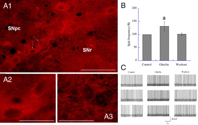
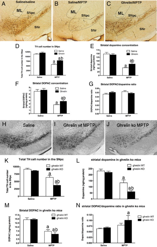
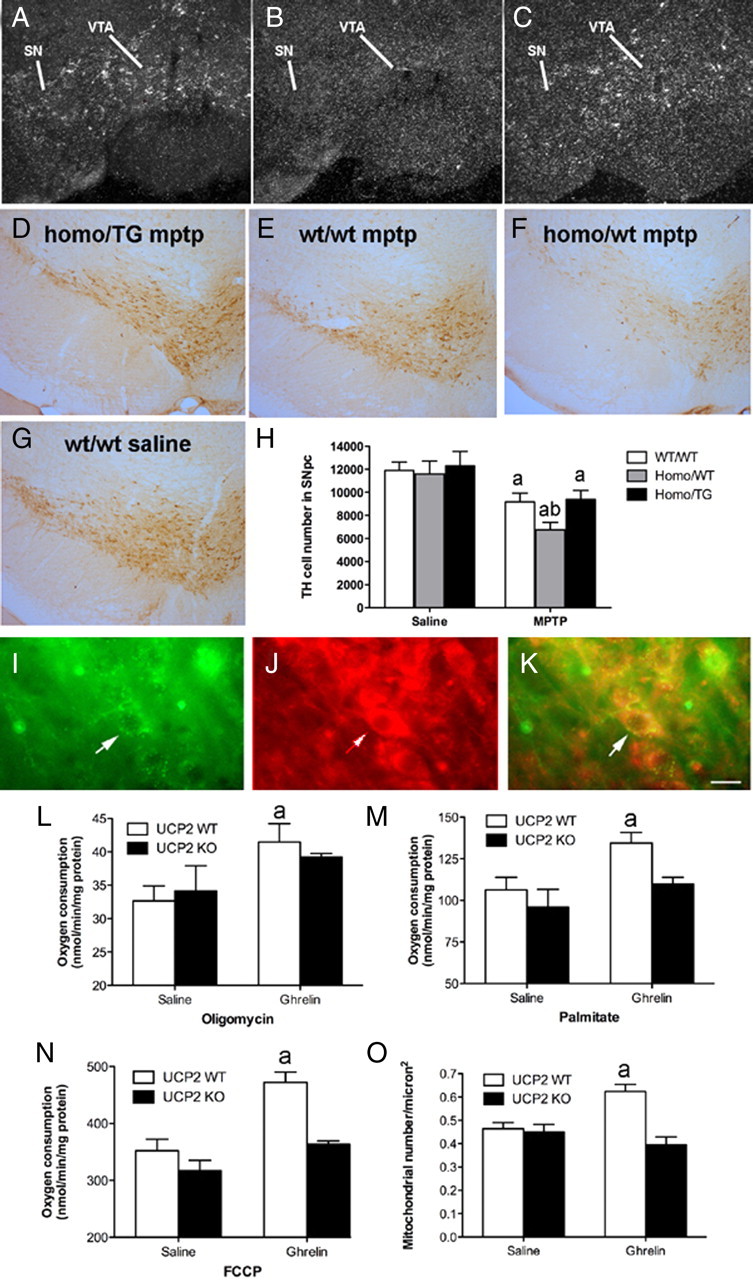
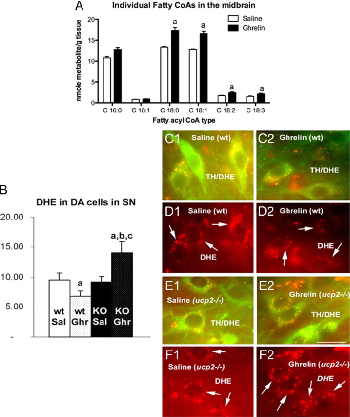
Ghrelin targets the hypothalamus to regulate food intake and adiposity. Endogenous ghrelin receptors [growth hormone secretagogue receptor (GHSR)] are also present in extrahypothalamic sites where they promote circuit activity associated with learning and memory, and reward seeking behavior. Here, we show that the substantia nigra pars compacta (SNpc), a brain region where dopamine (DA) cell degeneration leads to Parkinson's disease (PD), expresses GHSR. Ghrelin binds to SNpc cells, electrically activates SNpc DA neurons, increases tyrosine hydroxylase mRNA and increases DA concentration in the dorsal striatum. Exogenous ghrelin administration decreased SNpc DA cell loss and restricted striatal dopamine loss after 1-methyl-4-phenyl-1,2,5,6 tetrahydropyridine (MPTP) treatment. Genetic ablation of ghrelin or the ghrelin receptor (GHSR) increased SNpc DA cell loss and lowered striatal dopamine levels after MPTP treatment, an effect that was reversed by selective reactivation of GHSR in catecholaminergic neurons. Ghrelin-induced neuroprotection was dependent on the mitochondrial redox state via uncoupling protein 2 (UCP2)-dependent alterations in mitochondrial respiration, reactive oxygen species production, and biogenesis. Together, our data reveal that peripheral ghrelin plays an important role in the maintenance and protection of normal nigrostriatal dopamine function by activating UCP2-dependent mitochondrial mechanisms. These studies support ghrelin as a novel therapeutic strategy to combat neurodegeneration, loss of appetite and body weight associated with PD. Finally, we discuss the potential implications of these studies on the link between obesity and neurodegeneration.


Figures

Figure 1.
Figure 1.
Ghrelin targets SNpc DA neurons and regulates nigrostriatal DA function. A1, Low-power image showing ghrelin binding in the SNpc using biotinylated ghrelin. A2, High-power image of the neurons indicated by arrows in A1. A3, Absence of binding in the SNpc when unbiotinylated ghrelin is used. Both biotinylated ghrelin and unbiotinylated are used at 10 nmol. SNr, Substantia nigra reticulata. Scale bars: A1, 30 μm; A2, A3, 10 μm. B, Ghrelin significantly increased SNpc DA action potential frequency above baseline (n = 11 DA cells identified by characteristic I h currents). Data are presented as the mean ± SEM. aSignificant with respect to control, p < 0.05. Washout reduced ghrelin-elevated firing back to control levels. C, Representative action potential trace recordings from control, ghrelin, and washout conditions.
Figure 2.
Figure 2.
Ghrelin attenuates MPTP-induced nigrostriatal DA damage. A–C, Representative images showing MPTP-induced TH cell loss in the SNpc of saline/saline, saline/MPTP, or ghrelin/MPTP-treated mice. In each picture the SNpc, indicated by the black lines, is at the same approximate rostrocaudal extent. ML represents the medial lemniscus and SNr represents the substantia nigra reticulata. D, Unbiased stereological quantification of total TH cell number in the SNpc shows that ghrelin restricts MPTP-induced TH cell loss (n = 6). E, F, Ghrelin restricts dopamine loss (E) as well as DOPAC loss (F) in the striatum (n = 6). G, No difference in striatal DOPAC/dopamine ratio was observed (n = 6). aSignificant with respect to saline or ghrelin/saline-treated mice; bsignificantly increased compared with saline/MPTP-treated mice (p < 0.05). H–J, Lower-power images showing representative DA cell number in SNpc after saline (H, n = 10), MPTP to ghrelin wt (I, n = 8), or MPTP to ghrelin −/− mice (J, n = 9). Note the significant loss of DA neurons in ghrelin −/− mice treated with MPTP compared with ghrelin wt MPTP-treated mice. K, Stereological quantification shows that ghrelin −/− mice have significantly fewer surviving DA cells in the SNpc. L, Ghrelin −/− mice also exhibit reduced striatal DA content compared with wt after MPTP intoxication. M, Striatal DOPAC concentrations in ghrelin wt and ghrelin −/− mice did not differ. N, Striatal DOPAC/dopamine ratio was increased in ghrelin −/− mice but not ghrelin wt mice after MPTP. Data are presented as the mean ± SEM. aSignificant with respect to saline controls; bsignificant with respect to wt MPTP (p < 0.05).
Figure 3.
Figure 3.
Selective reactivation of GHSR TH neurons restricts MPTP-induced DA cell loss. A–C, Representative dark-field photomicrographs of in situ hybridization histochemistry experiments performed on mouse brains using a mouse GHSR-specific riboprobe. Shown are GHSR mRNA expression within the substantia nigra (SN) and ventral tegmental area (VTA) of wild-type mice (A), the lack of GHSR transcripts within the SN and VTA of ghsr−/− mice (B), and reactivated GHSR mRNA expression within the SN and VTA of “homo/TG” mice (C). D–G, Representative low-power images of TH staining in the SNpc after MPTP insults (n = 6 all groups) in all genotypes (E–G) compared with w/w saline (D). H, Unbiased stereological quantification of total TH cell number in the SNpc of mice treated with saline or MPTP.aSignificant with respect to saline-treated control mice. bSignificant with respect to w/w MPTP-treated mice (p < 0.05). I–O, Ghrelin promotes UCP2-dependent mitochondrial respiration and proliferation. I, Immunofluorescent image showing GHSR in the SNpc. J, UCP2 in the SNpc using β galactosidase, which is the protein encoded by the lacZ reporter gene. K, Merge showing that GHSRs and UCP2 are coexpressed in SNpc neurons. Scale bar, 1 μm. L, Ghrelin increases mitochondrial respiration after the addition of oligomycin in wt but not ucp2−/− mice (10 nmol, i.p.; n = 4). M, Elevated uncoupling activity in UCP2 wt but not ucp2−/− mice after ghrelin treatment as indicated by increased mitochondrial respiration after the addition of the free fatty acid palmitate (n = 4). N, FCCP increases total respiratory capacity in UCP2 wt but not ucp2−/− mice after ghrelin injection (n = 4, each sample represents 3 pooled midbrain dissections). O, Increased mitochondrial number in SNpc DA neurons after ghrelin injection 3 h earlier (10 nmol, n = 13–15). Data are presented as the mean ± SEM. aSignificant respect to wt saline.
Figure 4.
Figure 4.
Ghrelin regulates long-chain fatty CoAs and ROS in the midbrain. A, Individual LCFA CoAs measured 3 h after ghrelin injection (10 nmol, i.p.). aSignificant with respect to saline controls. B, Quantification of ROS in TH neurons of the SNpc using dihydroethidium (DHE) shows that ghrelin reduces TH ROS production in UCP2 wt mice but robustly increases ROS in ucp2−/− mice. This suggests that the inability of ghrelin to enhance UCP2 in ucp2−/− mice prevents the buffering of increased ROS produced by increased fatty acid oxidation. Data are presented as the mean ± SEM. aSignificant with respect to saline wt controls; b,csignificant with respect to saline UCP2 wt and −/−, respectively (n = 5, p < 0.05). C1, C2, Representative images showing dual-channel ROS production (red) in TH neurons (green) of the SNpc in saline- and ghrelin-treated UCP2 wt mice. D1, D2, Representative images showing single-channel ROS production in saline- and ghrelin-treated UCP2 wt mice. E1, E2, Representative images showing dual-channel ROS production (red) in TH neurons (green) of the SNpc in saline- and ghrelin-treated ucp2−/− mice. F1, F2, Representative images showing single-channel ROS production in saline- and ghrelin-treated ucp2−/− mice. Data are presented as the mean ± SEM. Scale bar (in E2) C–F, 10 μm.

Similar articles

Cited by

References

    1. Abbott RD, Ross GW, White LR, Nelson JS, Masaki KH, Tanner CM, Curb JD, Blanchette PL, Popper JS, Petrovitch H. Midlife adiposity and the future risk of Parkinson's disease. Neurology. 2002;59:1051–1057. - PubMed
    1. Abizaid A, Liu ZW, Andrews ZB, Shanabrough M, Borok E, Elsworth JD, Roth RH, Sleeman MW, Picciotto MR, Tschöp MH, Gao XB, Horvath TL. Ghrelin modulates the activity and synaptic input organization of midbrain dopamine neurons while promoting appetite. J Clin Invest. 2006;116:3229–3239. - PMC - PubMed
    1. Abou-Sleiman PM, Muqit MM, Wood NW. Expanding insights of mitochondrial dysfunction in Parkinson's disease. Nat Rev Neurosci. 2006;7:207–219. - PubMed
    1. Andrews ZB, Diano S, Horvath TL. Mitochondrial uncoupling proteins in the CNS: in support of function and survival. Nat Rev Neurosci. 2005a;6:829–840. - PubMed
    1. Andrews ZB, Horvath B, Barnstable CJ, Elsworth J, Yang L, Beal MF, Roth RH, Matthews RT, Horvath TL. Uncoupling protein-2 is critical for nigral dopamine cell survival in a mouse model of Parkinson's disease. J Neurosci. 2005b;25:184–191. Erratum (2005) 25(8):table of contents. - PMC - PubMed

Publication types

MeSH terms

LinkOut - more resources