. 2009 Nov 11;29(45):14108-19.
doi: 10.1523/JNEUROSCI.2055-09.2009.

Abeta immunotherapy protects morphology and survival of adult-born neurons in doubly transgenic APP/PS1 mice

Affiliations

Abeta immunotherapy protects morphology and survival of adult-born neurons in doubly transgenic APP/PS1 mice

Barbara Biscaro et al. J Neurosci. .

Abstract

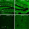
The hippocampus is heavily affected by progressive neurodegeneration and beta-amyloid pathology in Alzheimer's disease (AD). The hippocampus is also one of the few brain regions that generate new neurons throughout adulthood. Because hippocampal neurogenesis is regulated by both endogenous and environmental factors, we determined whether it benefits from therapeutic reduction of beta-amyloid peptide (Abeta)-related toxicity induced by passive Abeta immunotherapy. Abeta immunotherapy of 8-9-month-old mice expressing familial AD-causing mutations in the amyloid precursor protein and presenilin-1 genes with an antibody against Abeta decreased compact beta-amyloid plaque burden and promoted survival of newly born neurons in the hippocampal dentate gyrus. As these neurons matured, they exhibited longer dendrites with more complex arborization compared with newly born neurons in control-treated transgenic littermates. The newly born neurons showed signs of functional integration indicated by expression of the immediate-early gene Zif268 in response to exposure to a novel object. Abeta immunotherapy was associated with higher numbers of synaptophysin-positive synaptic boutons. Labeling dividing progenitor cells with a retroviral vector encoding green fluorescent protein (GFP) showed that Abeta immunotherapy restored the impaired dendritic branching, as well as the density of dendritic spines in new mature neurons. The presence of cellular prion protein (PrP(c)) on the dendrites of the GFP(+) newly born neurons is compatible with a putative role of PrP(c) in mediating Abeta-related toxicity in these cells. In addition, passive Abeta immunotherapy was accompanied by increased angiogenesis. Our data establish that passive Abeta immunotherapy can restore the morphological maturation of the newly formed neurons in the adult hippocampus and promote angiogenesis. These findings provide evidence for a role of Abeta immunotherapy in stimulating neurogenesis and angiogenesis in transgenic mouse models of AD, and they suggest the possibility that Abeta immunotherapy can recover neuronal and vascular functions in brains with beta-amyloidosis.


Figures

Figure 1.
Figure 1.
Aβ immunotherapy attenuated β-amyloid pathology. A, B, Immunohistochemical staining of brain tissue sections obtained from APP/PS1 mice treated with Aβ immunotherapy (α-Aβ) or a control antibody (ct ab) with a Cy3-conjugated antibody directed against mouse IgG revealed binding of the therapeutic antibody to β-amyloid plaques (B), an indication that the antibody had crossed the blood–brain barrier and reached the brain. CX, Cortex; CC, corpus callosum; LSI, lateral septal nuclei. C, D, Representative ThioS staining of compact β-amyloid plaques. CX, Cortex; DG, dentate gyrus; the dotted lines depict the corpus callosum (CC). There was a significantly lower area fraction of ThioS-positive β-amyloid plaques in APP/PS1 mice treated with Aβ immunotherapy (1.14 ± 0.14% area) compared with control antibody-treated mice (1.75 ± 0.16% area; p < 0.05; unpaired t test). Scale bars: (in A and B) 500 μm; (in C and D) 250 μm.
Figure 2.
Figure 2.
Aβ immunotherapy increased neurogenesis and dendritic arborization of newly born neurons in 11–12-month-old APP/PS1 mice. Aβ immunotherapy significantly increased numbers of mature BrdU+/NeuN+; this increase was accompanied by a trend for higher numbers of immature PSA-NCAM+ neurons (APP/PS1+ct ab: 306 ± 31 vs APP/PS1+α-Aβ: 452 ± 42 BrdU+/NeuN+ cells; p < 0.05; and APP/PS1+ct ab: 219 ± 79 vs APP/PS1+α-Aβ: 842 ± 275 PSA-NCAM+ cells; p = 0.06). A, B, Representative low-magnification images of PSA-NCAM+ neurons in the dentate gyrus of control-treated (A) and Aβ immunotherapy-treated (B) mice. Aβ immunotherapy significantly increased the number of dendrites of PSA-NCAM+ neurons in APP/PS1 mice and the length of dendrites of PSA-NCAM+ neurons in APP/PS1 mice (number of dendrites per cell: APP/PS1+ct ab: 2.45 ± 0.3 vs APP/PS1+α-Aβ: 4 ± 0.4; p < 0.01; and length of dendrites: APP/PS1+ct ab: 54.4 ± 7 vs APP/PS1+α-Aβ: 85.5 ± 10 μm; p < 0.01). C, D, High-magnification confocal details, including corresponding computer renderings resulting from quantitative NeuronJ analyses of the cells shown in the yellow squares in A and B, respectively, taken from comparable regions within the dentate gyrus. Scale bars: A, 60 μm; C, 20 μm. N = 40 cells were analyzed per group. Unpaired t test.
Figure 3.
Figure 3.
Newborn BrdU+ neurons expressed the activity-dependent immediate early gene Zif268 during Aβ immunotherapy as a sign of functional integration in pre-existing hippocampal synaptic circuits. Immunohistochemical stainings for the immediate-early gene product Zif268 (green), BrdU+ nuclei (red), NeuN+ neuronal nuclei (blue), overlay of the three channels (merge), and confocal orthogonal projections of merged Zif268 and BrdU images of selected granular neurons (ortho). A–D, Background expression of Zif268 in untreated APP/PS1 control mice without exposure to novel object stimulation: absent localization of Zif268 in nuclei of BrdU+ granular neurons. E–H, Presence of Zif268 in BrdU+ nuclei of granular neurons within the SGZ/GCL in response to novel object exposure in vehicle-treated APP/PS1 mice. I–M, Presence of Zif268 in BrdU+ nuclei of granular neurons within the SGZ/GCL during Aβ immunotherapy and in response to novel object exposure. Circles depict selected BrdU+ cells to facilitate their identification in the Zif268 staining. Scale bar: A, 10 μm.
Figure 4.
Figure 4.
Aβ immunotherapy increased the number of SYN-positive boutons in the ML of the hippocampus. A, B, SYN-positive synapses in the hippocampal outer ML from controls (ct ab; A) and Aβ immunotherapy-treated APP/PS1 mice (B). Dotted lines depict the area analyzed stereologically for quantification of SYN-positive boutons. Arrowheads show the presence of β-amyloid plaques. C, D, High magnification details of the yellow squares in A and B, respectively. The arrowhead in C depicts dystrophic boutons around a β-amyloid plaque. Stereological quantification of SYN-positive boutons in the ML revealed higher SYN levels in Aβ immunotherapy-treated mice (APP/PS1+ct ab: 1.4 ± 0.3 vs APP/PS1+α-Aβ: 2.8 ± 0.2 SYN+ boutons (× 106); p < 0.01; unpaired t test). Scale bars: A, 120 μm; C, 20 μm.
Figure 5.
Figure 5.
Aβ immunotherapy increased dendritic branching and spine densities of mature retrovirally labeled newly born neurons. A–C, Representative confocal pictures of GFP+ mature new neurons in the three groups of the study. Scale bar, 60 μm. D, Scholl analysis of new mature granule cells labeled with retrovirus expressing GFP revealed lower dendritic complexity in APP/PS1 mice compared with non-tg wild-type mice and restored dendritic complexity in APP/PS1 mice during Aβ immunotherapy compared with vehicle-treated APP/PS1 mice. The graph represents the mean number of intersections between dendrites and concentric radii, centered at the cell body, as a function of distance from the soma; n = 15 cells/group. Error bars represent SEM *p < 0.05 non-tg versus APP/PS1, and APP/PS1 versus APP/PS1 treated with α-Aβ. E, Representative high-magnification segments from dendrites of new mature neurons in non-tg, vehicle-treated APP/PS1, and α-Aβ-treated APP/PS1 mice. Scale bar, 10 μm. F–H, Computer-assisted classification of spines along 40 μm segments detected differences in the number of mushroom, long-thin, and stubby spines. F, Significant reduction of numbers of mushroom spines in APP/PS1 compared with non-tg mice and significant rescue during Aβ immunotherapy. G, Significant reduction of numbers of long-thin spines in APP/PS1 compared with non-tg mice and significant rescue during Aβ immunotherapy. H, Significant reduction of numbers of stubby spines in APP/PS1 compared with non-tg mice and a trend (p = 0.07) toward rescue during Aβ immunotherapy. N = 50 segments/group. Error bars represent SEM *p < 0.05, **p < 0.01, ***p < 0.001. ANOVA, followed by Bonferroni post hoc tests.
Figure 6.
Figure 6.
Aβ immunotherapy increased angiogenesis in the dentate gyrus. Stereological estimation of the number of blood vessels indicated significant increases in the number of blood vessels in APP/PS1 mice during Aβ immunotherapy (APP/PS1+ct ab: 5991 ± 384 vs APP/PS1+α-Aβ: 8978 ± 766 blood vessels; p < 0.01; unpaired t test). A, B, Confocal images of ThioS+-Aβ-deposits along the wall of blood vessels in the cortex. C, Disruption of Glut1 expression in the presence of congophilic amyloid angiopathy. Arrow depicts colocalization of Aβ with Glut1, excluding the possibility that Glut1 was present but not detected because of epitope-masking by β-amyloid. D, Glut1-stained blood vessels (blue) and BrdU (red) in the dentate gyrus: many BrdU+ cells are located in the vicinity of blood vessels. Glut1 receptor is expressed at highest levels in the endothelial cells of barrier tissues such as the blood-brain barrier. Dotted lines in A depict SGZ. Scale bars: A, 40 μm; D, 70 μm.
Figure 7.
Figure 7.
Expression of cellular prion protein (PrPc) on the dendrites of newly born neurons. A, Colocalization analysis of PrPc (red) with the GFP+ dendrites (green) and overlay (yellow) by ColocImaris software. Inset, Maximum projection of this image stack. B, Three-dimensional reconstruction using Imaris Surpass of both PrPc and GFP channels shows the broad presence of PrPc on brain cells along with its colocalization on the dendrites of newly born neurons. Scale bar, 4 μm.

Similar articles

Cited by

References

    1. Abdipranoto A, Wu S, Stayte S, Vissel B. The role of neurogenesis in neurodegenerative diseases and its implications for therapeutic development. CNS Neurol Disord Drug Targets. 2008;7:187–210. - PubMed
    1. Altman J, Das GD. Autoradiographic and histological evidence of postnatal hippocampal neurogenesis in rats. J Comp Neurol. 1965;124:319–335. - PubMed
    1. Arvidsson A, Collin T, Kirik D, Kokaia Z, Lindvall O. Neuronal replacement from endogenous precursors in the adult brain after stroke. Nat Med. 2002;8:963–970. - PubMed
    1. Bard F, Cannon C, Barbour R, Burke RL, Games D, Grajeda H, Guido T, Hu K, Huang J, Johnson-Wood K, Khan K, Kholodenko D, Lee M, Lieberburg I, Motter R, Nguyen M, Soriano F, Vasquez N, Weiss K, Welch B, Seubert P, Schenk D, Yednock T. Peripherally administered antibodies against amyloid β-peptide enter the central nervous system and reduce pathology in a mouse model of Alzheimer disease. Nat Med. 2000;6:916–919. - PubMed
    1. Becker M, Lavie V, Solomon B. Stimulation of endogenous neurogenesis by anti-EFRH immunization in a transgenic mouse model of Alzheimer's disease. Proc Natl Acad Sci U S A. 2007;104:1691–1696. - PMC - PubMed

Publication types

MeSH terms