. 2009 Nov 11;29(45):14120-6.
doi: 10.1523/JNEUROSCI.1726-09.2009.

Individual calcium syntillas do not trigger spontaneous exocytosis from nerve terminals of the neurohypophysis

Affiliations

Individual calcium syntillas do not trigger spontaneous exocytosis from nerve terminals of the neurohypophysis

James M McNally et al. J Neurosci. .

Abstract

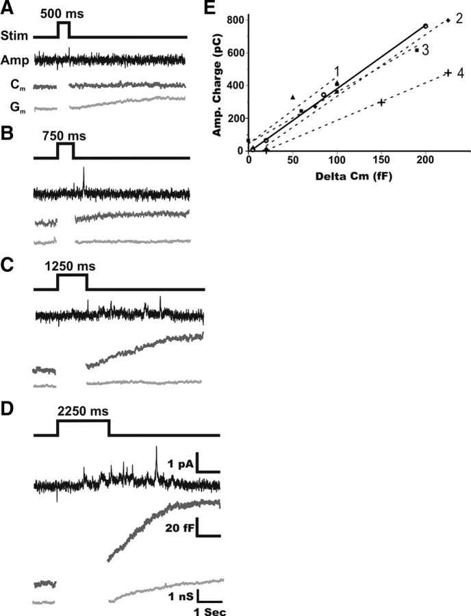
Recently, highly localized Ca(2+) release events, similar to Ca(2+) sparks in muscle, have been observed in neuronal preparations. Specifically, in murine neurohypophysial terminals (NHT), these events, termed Ca(2+) syntillas, emanate from a ryanodine-sensitive intracellular Ca(2+) pool and increase in frequency with depolarization in the absence of Ca(2+) influx. Despite such knowledge of the nature of these Ca(2+) release events, their physiological role in this system has yet to be defined. Such localized Ca(2+) release events, if they occur in the precise location of the final exocytotic event(s), may directly trigger exocytosis. However, directly addressing this hypothesis has not been possible, since no method capable of visualizing individual release events in these CNS terminals has been available. Here, we have adapted an amperometric method for studying vesicle fusion to this system which relies on loading the secretory granules with the false transmitter dopamine, thus allowing, for the first time, the recording of individual exocytotic events from peptidergic NHT. Simultaneous use of this technique along with high-speed Ca(2+) imaging has enabled us to establish that spontaneous neuropeptide release and Ca(2+) syntillas do not display any observable temporal or spatial correlation, confirming similar findings in chromaffin cells. Although these results indicate that syntillas do not play a direct role in eliciting spontaneous release, they do not rule out indirect modulatory effects of syntillas on secretion.


Figures

Figure 1.
Figure 1.
False transmitter loading of whole-cell patched NHT allows detection of spontaneous amperometric events similar to those seen in chromaffin cells. A, Amperometric recording from an isolated murine adrenal chromaffin cell showing spontaneous release events. B, Amperometric recording performed immediately after whole-cell patch of an NHT voltage clamped at −80 mV shows that no amperometric activity is detectable in a non-DA-loaded terminal. After several minutes, allowing DA to dialyze into the terminal from the patch pipette, spontaneous amperometric release events can be observed. C, Amperometric events recorded from both chromaffin cells and false transmitter-loaded terminals appear very similar in nature, with the exception of their size. Prespike feet, a hallmark of amperometric recording of quantal release events, can be seen in a number of events (right) recorded from loaded NHTs.
Figure 2.
Figure 2.
Amperometric activity in false transmitter-loaded terminals correlates with exocytotic activity observed using membrane capacitance recording. A–D, Simultaneous amperometric (Amp) and membrane capacitance (Cm) recordings taken from a representative false transmitter-loaded terminal. The terminal was given a series of depolarizing stimuli (−80 to 0 mV) increasing in duration from A to D. Both the amount of amperometric activity and the evoked change in membrane capacitance increased with longer durations of depolarizing stimuli. Conductance traces (Gm) for each recording are also displayed. Of note, in A and D, an increase in conductance can be observed after cessation of stimulus. As changes in conductance do not correlate with the changes in capacitance, it does not appear that Gm changes had an effect on capacitance recordings. E, Plotting the total amperometric charge versus the evoked change in capacitance (delta Cm) reveals a strong linear correlation. Here, the total change in capacitance was measured as the difference between the capacitance just before the stimulus and the maximum observed capacitance after the stimulus (including asynchronous activity). Total amperometric charge was calculated from the integral of the amperometric current trace over the same time period found between the onset of stimulus and peak capacitance value. Data obtained from the terminal shown (open circles) was representative of the correlation seen between amperometric and membrane capacitance recordings taken from a number of individual terminals (solid black line; r2 = 0.99). Dashed lines 1–4 show four additional examples of terminals where at least three different duration depolarizing stimuli where applied. The comparative amperometric and capacitance data (triangles, diamonds, squares, cross, respectively) as well as the linear fit (r2 = 0.89, 0.97, 0.99, 0.99, respectively) are plotted.
Figure 3.
Figure 3.
Calcium syntillas do not trigger spontaneous release events. In false transmitter-loaded terminals, voltage clamped at −60 mV, Ca2+ imaging of syntillas and amperometric recording of exocytotic events can be performed simultaneously. Top, Leftmost image shows positioning of the CFE relative to a whole-cell patched terminal loaded with the false transmitter, dopamine, and the Ca2+ indicator dye, fluo-3. Images 1–5 represent the image frames taken immediately before the appearance of the syntilla and the following four frames, respectively. The syntilla is observed to occur precisely in the area where the CFE is positioned. Bottom, Concurrent records of fluorescence in the region of the terminal where the syntilla occurs (red trace) and amperometric current (black trace) show that there are no amperometric events immediately correlated with this syntilla, despite the fact that this syntilla represents the release of ∼400,000 Ca2+ ions [signal mass (SM) = 66.4 × 10⁻²⁰ mol Ca2+] in the precise area where the CFE is located. The numbers on the fluorescence trace correspond to the numbered images above.
Figure 4.
Figure 4.
There is no temporal correlation between syntillas and spontaneous release events. Syntillas observed occurring in the region of the terminal where a CFE electrode had been placed were plotted based on their signal mass (SM). Filled triangles represent syntillas occurring near the CFE where long duration amperometric recording was performed before and after the syntilla. For these syntillas, the closest spontaneous amperometric events occurring both before and after the syntilla (black circles) have been plotted. Hollow triangles represent syntillas where amperometric recording was performed only during Ca2+ imaging. A, Plot of syntillas versus amperometric events shows that there is no apparent temporal correlation on the scale predicted be required if syntillas were directly responsible for eliciting exocytosis (see Results). B, Expanding the time span of the previous graph to include the nearest preceding and following amperometric event (filled triangles only) still does not provide any evidence of temporal correlation.
Figure 5.
Figure 5.
Spontaneous amperometric release events do not trigger calcium syntillas. As in Figure 3, Ca2+ imaging of syntillas and amperometric recording of exocytotic events were performed simultaneously in false transmitter-loaded terminals voltage clamped at −60 mV. Top, Leftmost image shows positioning of CFE relative to a whole-cell patch-clamped terminal. Images 1–5 represent the image frames taken during the occurrence of the spontaneous false transmitter release event. Bottom, Concurrent records of fluorescence in the region of terminal where the CFE was positioned (red trace) and amperometric current (black trace) shows that there are no Ca2+ release events occurring near the CFE when the spontaneous release event is detected. The numbers on the fluorescence trace correspond to the temporal position of the numbered images above.

Similar articles

Cited by

References

    1. Becherer U, Moser T, Stühmer W, Oheim M. Calcium regulates exocytosis at the level of single vesicles. Nat Neurosci. 2003;6:846–853. - PubMed
    1. Berridge MJ. Calcium microdomains: organization and function. Cell Calcium. 2006;40:405–412. - PubMed
    1. Cazalis M, Dayanithi G, Nordmann JJ. Hormone release from isolated nerve endings of the rat neurohypophysis. J Physiol. 1987;390:55–70. - PMC - PubMed
    1. Chen P, Gillis KD. The noise of membrane capacitance measurements in the whole-cell recording configuration. Biophys J. 2000;79:2162–2170. - PMC - PubMed
    1. Chow RH, von Rüden L, Neher E. Delay in vesicle fusion revealed by electrochemical monitoring of single secretory events in adrenal chromaffin cells. Nature. 1992;356:60–63. - PubMed

Publication types

LinkOut - more resources