. 2009 Nov 11;29(45):14199-210.
doi: 10.1523/JNEUROSCI.2873-09.2009.

Overexpressing temperature-sensitive dynamin decelerates phototransduction and bundles microtubules in Drosophila photoreceptors

Affiliations

Overexpressing temperature-sensitive dynamin decelerates phototransduction and bundles microtubules in Drosophila photoreceptors

Paloma T Gonzalez-Bellido et al. J Neurosci. .

Abstract

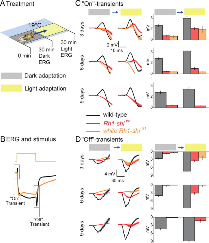
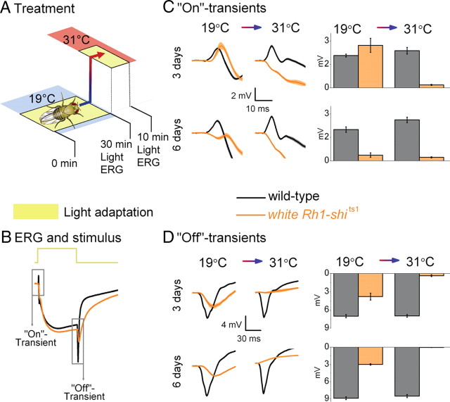
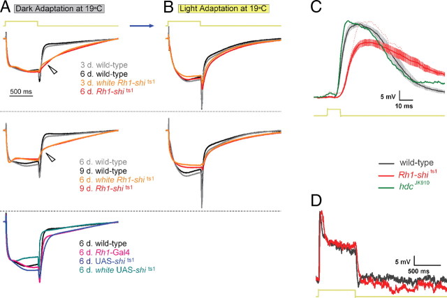
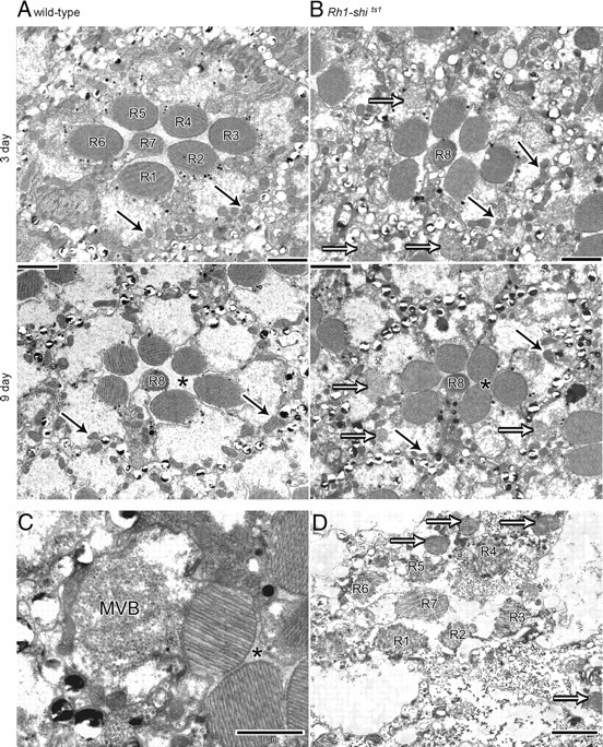
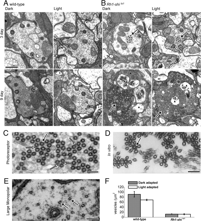
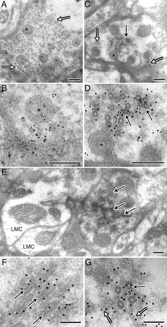
shibire(ts1), a temperature-sensitive mutation of the Drosophila gene encoding a Dynamin orthologue, blocks vesicle endocytosis and thus synaptic transmission, at elevated, or restrictive temperatures. By targeted Gal4 expression, UAS-shibire(ts1) has been used to dissect neuronal circuits. We investigated the effects of UAS-shibire(ts1) overexpression in Drosophila photoreceptors at permissive (19 degrees C) and restrictive (31 degrees C) temperatures. At 19 degrees C, overexpression of UAS-shi(ts1) causes decelerated phototransduction and reduced neurotransmitter release. This phenotype is exacerbated with dark adaptation, age and in white mutants. Photoreceptors overexpressing UAS-shibire(ts1) contain terminals with widespread vacuolated mitochondria, reduced numbers of vesicles and bundled microtubules. Immuno-electron microscopy reveals that the latter are dynamin coated. Further, the microtubule phenotype is not restricted to photoreceptors, as UAS-shibire(ts1) overexpression in lamina cells also bundles microtubules. We conclude that dynamin has multiple functions that are interrupted by UAS-shibire(ts1) overexpression in Drosophila photoreceptors, destabilizing their neural communication irreversibly at previously reported permissive temperatures.


Figures

Figure 1.
Figure 1.
Rh1-shits1 photoreceptor output depends on light history at 19°C. A, Dark adaptation (30 min) followed by light adaptation (30 min) at 19°C. B, ERGs were recorded (700 ms stimulus) after each adaptation. Traces are from dark-adapted flies and 6 d posteclosion. C, D, The amplitude of “On”-transients (C) and “Off”-transients (D) quantified for wild type (black), Rh1-shits1 (red), and white Rh1-shits1 (orange). Mean ± SEM is shown in traces and histograms with n = 6 for each group. Age refers to days posteclosion.
Figure 2.
Figure 2.
Temperature shift in light-adapted Rh1-shits1 photoreceptors. A, Light adaptation (30 min) at 19°C before light adaptation (10 min) at 31°C. B, ERGs were recorded (700 ms stimulus) after each adaptation. Traces are from light-adapted flies at 19°C and 6 d posteclosion. C, D, The amplitude of “On”-transients (C) and “Off”-transients (D) quantified for wild type (black) and white Rh1-shits1 (orange). Mean ± SEM is shown in traces and histograms with n = 6 for each group. Age refers to days posteclosion.
Figure 3.
Figure 3.
Neurotransmission from transgenic controls. A, Dark adaptation (30 min) at 19°C before dark adaptation (10 min) at 31°C. B, ERGs were recorded (700 ms stimulus) after each adaptation. Traces are from dark-adapted flies at 19°C and 6 d posteclosion. The amplitude of “On”-transients (C) and “Off”-transients (D) quantified for wild-type (black) and homozygous transgenic controls, Rh1-Gal4 (pink), UAS-shits1 (dark blue), and white UAS-shits1 (blue-green). Mean ± SEM is shown in traces and histograms with n = 6 for each group. Age refers to days posteclosion.
Figure 4.
Figure 4.
Assessment of Gal4 and UAS insertions. A, Dark adaptation (30 min) at 19°C before dark adaptation (10 min) at 31°C. ERGs were recorded (700 ms stimulus) after each adaptation. Traces are from dark-adapted flies at 19°C and 6 d posteclosion. Genotypes range in number of Rh1-Gal4 and UAS-shits1 insertions. B, C, The amplitude of “On”-transients (B) and “Off”-transients (C) quantified for wild type (black), white/+; Rh1-Gal4/+; UAS-shits1/+ (blue), white; Rh1-Gal4/+; UAS-shits1 (green), and white/+; Rh1-Gal4; UAS-shits1/+ (pink). D, E, “On”-transients (D) and “Off”-transients (E) from white/+; Rh1-Gal4; UAS-shits1/+ and wild-type flies raised at 16°C up to 3 d posteclosion and thereafter shifted to 18°C. Mean ± SEM is shown in traces and histograms with n = 6 for each group. Age refers to days posteclosion.
Figure 5.
Figure 5.
Rh1-shits1 photoreceptors show decelerated voltage responses to light. Mean normalized ERGs (n = 6) to 700 ms stimulus, taken from Figures 1 and 3 after dark adaptation (A) and after light adaptation (B). Wild type is plotted for young (gray) and older (black) flies. white Rh1-shits1 (orange) and Rh1-shits1 (red) at 3 and 6 d posteclosion, respectively, have been plotted together. Similarly, 6 and 9 d posteclosion are shown together. Wild-type (black) and homozygous transgenic controls; Rh1-Gal4 (pink), UAS-shits1 (dark blue) and white UAS-shits1 (blue-green) have been plotted together (bottom). Arrows indicate differences in response waveforms between overexpressed UAS-shits1 and wild type. Age refers to days posteclosion. C, Intracellular recordings of 3 d posteclosion wild-type (black) and Rh1-shits1 (red) photoreceptors. Mean ± SEM shown in traces (n = 9). When Rh1-shits1 displayed the same light response amplitude (red dotted line) as wild type, the dynamics were as slow as the rest of the cells. The histamine mutant hdcJK910 (green) does not show such delay in its response. D, Representative intracellular recordings of wild type (black) and Rh1-shits1 (red) for a 700 ms light stimulus.
Figure 6.
Figure 6.
Overexpression of UAS-shits1 changes retinal morphology at 19°C. EM cross-sections of 3 and 9 d posteclosion wild-type (A) and Rh1-shits1 (B) retinas. Scale = 2 μm. C, MVBs present in Rh1-shits1 photoreceptor. Scale = 1 μm. D, Rh1-shits1 retina, raised at 25°C, with photoreceptor R7 in a healthy state, demonstrating that Rh1-Gal4 overexpresses UAS-shits1 only in photoreceptors R1–R6. Scale = 2 μm. Mitochondria (solid arrows), Multivesicular bodies (open arrows), inter-photoreceptor space (asterisk).
Figure 7.
Figure 7.
Overexpression of UAS-shits1 changes photoreceptor terminal morphology at 19°C. A, B, EM cross-sections of 3 and 9 d posteclosion wild-type (A) and Rh1-shits1 (B) terminals, reared at 18°C. Flies were exposed to 30 min of darkness (Dark) or to 30 min of darkness followed by 30 min of light (Light) at 18°C. Scale, 500 nm. Mitochondria (m), coated microtubules (solid arrows), capitate projections (asterisk), and apoptotic mitochondria profiles (black triangle) are shown. C, High-magnification image of coated microtubules with cross-bridges (open arrow). D, In vitro coated microtubules, [Shpetner and Vallee (1989); their Fig. 6B, reprinted with permission]. E, Image of LMC wild-type microtubules. Scale (C–E), 100 nm; microtubules (solid arrow). F, Number of vesicles per square micrometer for 3 d posteclosion wild type (dark-adapted; 91.40 ± 11.02, light-adapted; 68.86 ± 3.07) and Rh1-shits1 (dark-adapted; 12.70 ± 0.78, light-adapted; 11.70 ± 2.26) terminals. Mean ± SEM is given.
Figure 8.
Figure 8.
Immunogold labeling confirms that the microtubule coat is dynamin. A, Wild-type terminal showing 10 nm gold immunolabeling in proximity to the membrane, indicated by open arrows and capitate projections (asterisk). B, High magnification of A. C, Photoreceptor terminal from Rh1-shits1, which in addition to labeling near capitate projections (open arrows), has labeling in the cytoplasm near coated microtubules (solid arrow). D, High magnification of C. E, Terminal from Rh1-shits1 showing labeling of oblique coated microtubules in the cytoplasm. Note that adjacent LMC profiles do not have cytoplasmic labeling, thus confirming the specificity of gold labeling to overexpressed shits1 dynamin. Scale (A–E), 200 nm. F, G, High magnification of labeled oblique (F) and cross-section of coated (G) microtubules (solid arrow) showing that labeling extends to filaments adjacent (open arrow). Scale (F–G), 100 nm.
Figure 9.
Figure 9.
Microtubule coat is present in other neurons when UAS-shits1 is overexpressed. A, MJ85b-shits1 lamina cartridge showing coated microtubules (arrows) in lamina cells and not in photoreceptor terminals (R). Scale, 1 μm. B, Close-up of coated microtubules in a lamina cell. Scale, 0.2 μm. C, Lamina cell spine packed with coated microtubules. Scale, 0.5 μm.

Similar articles

Cited by

References

    1. Acharya U, Patel S, Koundakjian E, Nagashima K, Han X, Acharya JK. Modulating sphingolipid biosynthetic pathway rescues photoreceptor degeneration. Science. 2003;299:1740–1743. - PubMed
    1. Bähner M, Frechter S, Da Silva N, Minke B, Paulsen R, Huber A. Light-regulated subcellular translocation of Drosophila TRPL channels induces long-term adaptation and modifies the light-induced current. Neuron. 2002;34:83–93. - PubMed
    1. Beramendi A, Peron S, Casanova G, Reggiani C, Cantera R. Neuromuscular junction in abdominal muscles of Drosophila melanogaster during adulthood and aging. J Comp Neurol. 2007;501:498–508. - PubMed
    1. Borycz J, Borycz JA, Kubów A, Lloyd V, Meinertzhagen IA. Drosophila ABC transporter mutants white, brown and scarlet have altered contents and distribution of biogenic amines in the brain. J Exp Biol. 2008;211:3454–3466. - PubMed
    1. Brand AH, Perrimon N. Targeted gene expression as a means of altering cell fates and generating dominant phenotypes. Development. 1993;118:401–415. - PubMed

Publication types

MeSH terms

LinkOut - more resources