. 2009 Nov 11;29(45):14257-64.
doi: 10.1523/JNEUROSCI.4403-09.2009.

Polar residues in the second transmembrane domain of the rat P2X2 receptor that affect spontaneous gating, unitary conductance, and rectification

Affiliations

Polar residues in the second transmembrane domain of the rat P2X2 receptor that affect spontaneous gating, unitary conductance, and rectification

Lishuang Cao et al. J Neurosci. .

Abstract

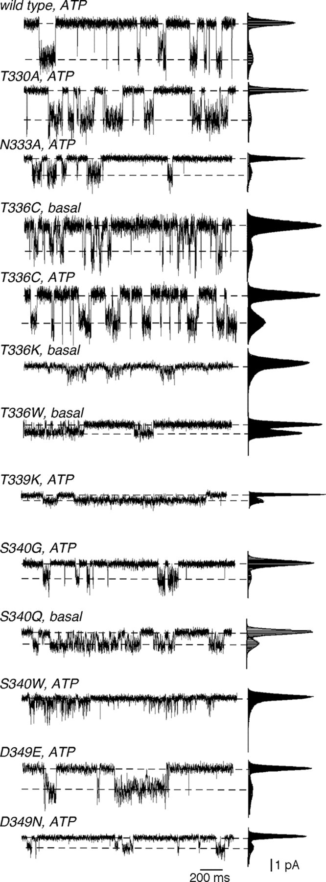
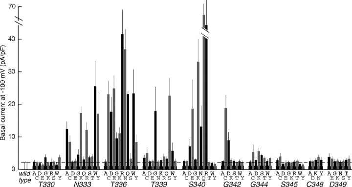
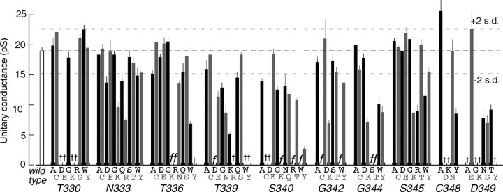
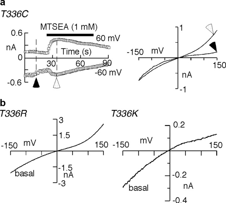
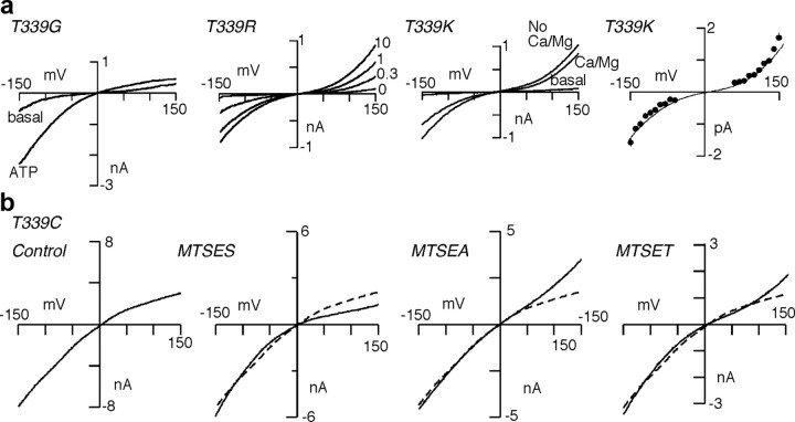
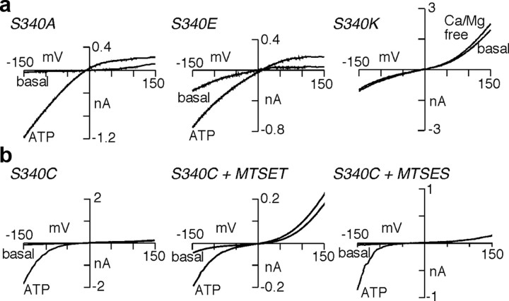
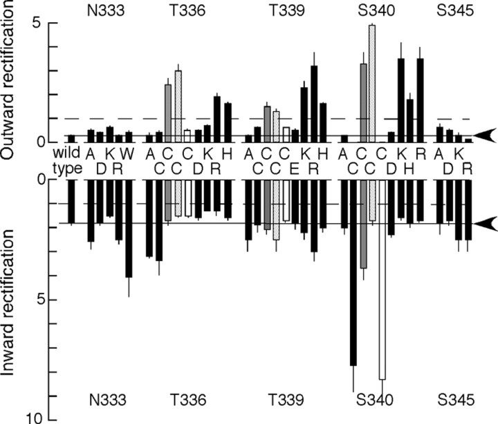
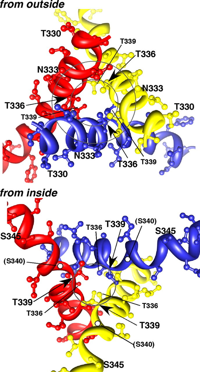
Membrane ion channels activated by extracellular ATP (P2X receptors) are widely distributed in the nervous system. Their molecular architecture is fundamentally distinct from that of the nicotinic or glutamate receptor families. We have measured single-channel currents, spontaneous gating, and rectification of rat P2X2 receptor in which polar and charged residues of the second transmembrane domain (TM2) were systematically probed by mutagenesis. The results suggest that Asn(333) and Asp(349) lie respectively in external and internal vestibules. Substitutions at Asn(333), Thr(336), and Ser(340) were particularly likely to cause spontaneously active channels. At Thr(336), Thr(339), and Ser(340), the introduction of positive charge (Arg, Lys, or His, or Cys followed by treatment with 2-aminoethyl methanethiosulphonate) greatly enhanced outward currents, suggesting that side-chains of these three residues are exposed in the permeation pathway of the open channel. These functional findings are interpreted in the context of the recently reported 3.1 A crystal structure of the zebrafish P2X4.1 receptor in the closed state. They imply that the gate is formed by residues Asn(333) to Thr(339) and that channel opening involves a counter-clockwise rotation and separation of the TM2 helices.


Figures

Figure 1.
Figure 1.
Sequences of the second transmembrane domain of P2X receptors. Sequence comparisons among six rat P2X receptors, four invertebrate receptors, and one zebrafish receptor, all of which form functional ATP-gated channels. 1, P2X1 (accession P47824); 2, P2X2 (P49653); 3, P2X3 (P49654); 4, P2X4 (P51577); 5, P2X5 (51578); 7, P2X7 (Q64663); Sm, Schistosoma mansoni (CAH04147); Dd, Dictyostelium discoideum (ABS88293); Ot, Ostreococcus tauri (CAL54489); Mb, Monosiga brevicollis (XP_001743535); zf4, Zebrafish P2X4.1 (AF317643). Residues mutated in this study are circled.
Figure 2.
Figure 2.
Examples of unitary currents in mutated P2X2 receptors. In each trace, outside-out recording is shown to the left. At the right are shown all-points histograms from longer periods of recording (at least 30 opening events). The vertical axis is the current level with the same scale as the current traces to the left. The horizontal axis is the point count, with a scale range for the open state of 1000–10,000 points. The distribution was fitted by the sum of two Gaussians, and the peaks of these fits were used to estimate unitary current amplitudes. Some examples show spontaneously opening channels in the absence of ATP (basal). Others show activity evoked by adding ATP [concentrations were as follows (in μm): wild type, 0.3; T330A, 1; N333A, 0.3; T336C, 0.3; T339K, 0.3; S340G, 1; S340W, 3; D349E, 1; D349N, 10].
Figure 3.
Figure 3.
Standing inward currents observed for P2X2 receptors with amino acid substitutions in TM2. Ordinate shows mean holding current (at −100 mV) in cells expressing the point mutation shown on the abscissa. Broken line is wild-type level. Bars are SEM for 5–11 observations, except 33 for wild type.
Figure 4.
Figure 4.
Unitary conductance of ATP-evoked currents in excised patches, for P2X channels with single amino substitutions in TM2. f, Too flickery for unitary currents to be resolved. †, No current or very small current evoked by ATP (excised patch or whole-cell recording). In those cases where spontaneously occurring unitary currents were resolved (Fig. 2), the application of ATP increased open probability but did not obviously change unitary current amplitudes. In other cases (for N333K, N333R, T336D, T336E, T336N, T336Q, T336W, S340E, S340K, S340N, S3430Q), where there was no response to ATP, the unitary current amplitudes are from spontaneously occurring openings. Conductance was estimated from all-points histograms at −100 mV. Bars are SEM from 5 to 12 patches in each case.
Figure 5.
Figure 5.
Rectification introduced by substitutions at Thr336. a, Left, Time course of currents measured at −60 mV and +60 mV in T336C. Right, Current–voltage relation from voltage ramps applied at the times indicated by the arrowheads. The basal current of T336C shows similar inward rectification to wild-type channels (filled arrowhead), but after MTSEA (1 mm, solid bar), the current shows marked outward rectification at positive potentials (open arrowhead). b, Left, T336R shows large basal current activity, with marked outward rectification. Right, Outward rectification is much less in T336K.
Figure 6.
Figure 6.
Rectification introduced by substitutions at Thr339. a, Left, T339G shows substantial basal current, and this is increased by ATP (0.3 μm). Inward rectification is similar to that seen in wild-type channels. Center-left, T339R shows no basal current (trace labeled 0), but ATP (concentrations indicated, μm)-evoked currents showing marked outward rectification at positive potentials. Center-right, T339K shows no basal current, but ATP (10 μm)-evoked currents showing marked outward rectification at positive potentials. This rectification was unaffected by removal of calcium and magnesium from the external solution (No Ca/Mg). Right, Unitary currents in T339K show strong rectification both for inward and outward currents. Points are mean amplitudes from seven patches; SEM is less than symbol size. Solid line is scaled whole-cell I–V relation. b, Left, T339C currents evoked by ATP (3 μm in each case) show inward rectification at positive potentials similar to see in wild-type receptors. Center-left, This inward rectification is more marked after addition of MTSES (1 mm, 5 min). Center-right and right, The rectification becomes outward after treatment with MTSEA (1 mm, 5 min) or MTSET (1 mm, 5 min). The broken line in the right three panels is the scaled I–V relation from the left panel.
Figure 7.
Figure 7.
Rectification introduced by substitutions at Ser340. a, Left, S340A was similar to wild-type channels; there was minimal basal current, and the current evoked by ATP (3 μm) showed typical inward rectification. Center, S340E showed a small basal current and additional current evoked by ATP (30 μm). Right, S340K shows outward rectification. This is not different in no extracellular calcium or magnesium. b, Left, S340C shows no basal activity, and ATP elicits a current with typical inward rectification. Center, After addition of MTSET, both basal and ATP-evoked currents show marked outward rectification. Right, After addition of MTSES, neither basal nor ATP-evoked current shows outward rectification. ATP (30 μm) in each case.
Figure 8.
Figure 8.
Summary results for mutations causing outward rectification. At Thr336, Thr339, and Ser340, positively charged residues or positively charged MTS compounds introduced marked outward rectification. Rectification is expressed as the additional current observed at +150 mV (outward rectification, top) or −150 mV (inward rectification, bottom) compared with that which would be observed by linear extrapolation of the current–voltage relation from the range −5 to +5 mV; therefore, unity (broken line) indicates no rectification. Solid line and arrowheads indicate rectification in wild-type P2X receptors. Filled gray columns, Cysteine substitution after addition of MTSEA. Stippled columns, Cysteine substitution after addition of MTSET. Open columns, Cysteine substitution after addition of MTSES. ATP concentrations were as follows (in μm): wild-type, 3; N333A, 1; N333D, 3; N333K, 0; N333R, 0; N333W, 1; T336A, 3; T336C, 3; T336K, 0; T336R, 0; T336H, 10; T339A, 1; T339E, 3; T339C, 3; T339K, 1; T339R, 1; T339H, 3; S340A, 10; S340C, 30; S340D, 0; S340H, 0; S340K, 0; S340R, 0; S345A, 10; S345D, 10; S345K, 10; S345R, 10. Where the concentration is indicated as zero, the rectification of the “standing” current introduced by the substitution is presented.
Figure 9.
Figure 9.
Model of three TM2 helices to illustrate positions of key residues. Viewed from the outside, the circles pass through the Cα atoms of Asn333 (outer circle) and Thr336 (inner circle) from each chain. Viewed from the inside, the circle passes through the Cα atoms of Thr339. The structure shown is a threaded model of rat P2X2 generated using a default script in Modeler9v6 from the zebrafish P2X4 receptor in the closed state (pdb 3H9V) (Kawate et al., 2009) and indicating side-chains from Asn333 to Ser340 only. The side-chain of Ser340 is not seen, but the position of Ser340 Cα is indicated (white dots). The results indicate that Ser340 is exposed in the open channel permeation pathway, which implies that the three TM2 helices twist counter-clockwise during channel opening.

Similar articles

Cited by

References

    1. Brake AJ, Wagenbach MJ, Julius D. New structural motif for ligand-gated ion channels defined by an ionotropic ATP receptor. Nature. 1994;371:519–523. - PubMed
    1. Cao L, Young MT, Broomhead HE, Fountain SJ, North RA. Thr339-to-serine substitution in rat P2X2 receptor second transmembrane domain causes constitutive opening and indicates a gating role for Lys308. J Neurosci. 2007;27:12916–12923. - PMC - PubMed
    1. Clyne JD, Brown TC, Hume RI. Expression level dependent changes in the properties of P2X2 receptors. Neuropharmacology. 2003;44:403–412. - PubMed
    1. Ding S, Sachs F. Single channel properties of P2X2 purinoceptors. J Gen Physiol. 1999a;113:695–720. - PMC - PubMed
    1. Ding S, Sachs F. Ion permeation and block of P2X2 purinoceptors: single channel recordings. J Membr Biol. 1999b;172:215–223. - PubMed

Publication types

LinkOut - more resources