. 2009 Nov 11;29(45):14287-98.
doi: 10.1523/JNEUROSCI.3976-09.2009.

Trafficking of membrane proteins to cone but not rod outer segments is dependent on heterotrimeric kinesin-II

Affiliations

Trafficking of membrane proteins to cone but not rod outer segments is dependent on heterotrimeric kinesin-II

Prachee Avasthi et al. J Neurosci. .

Abstract

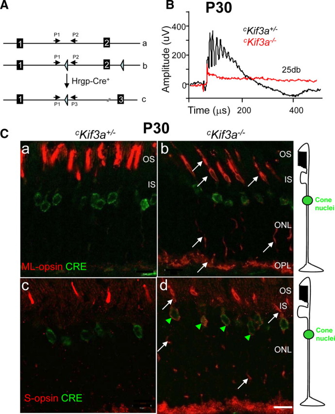
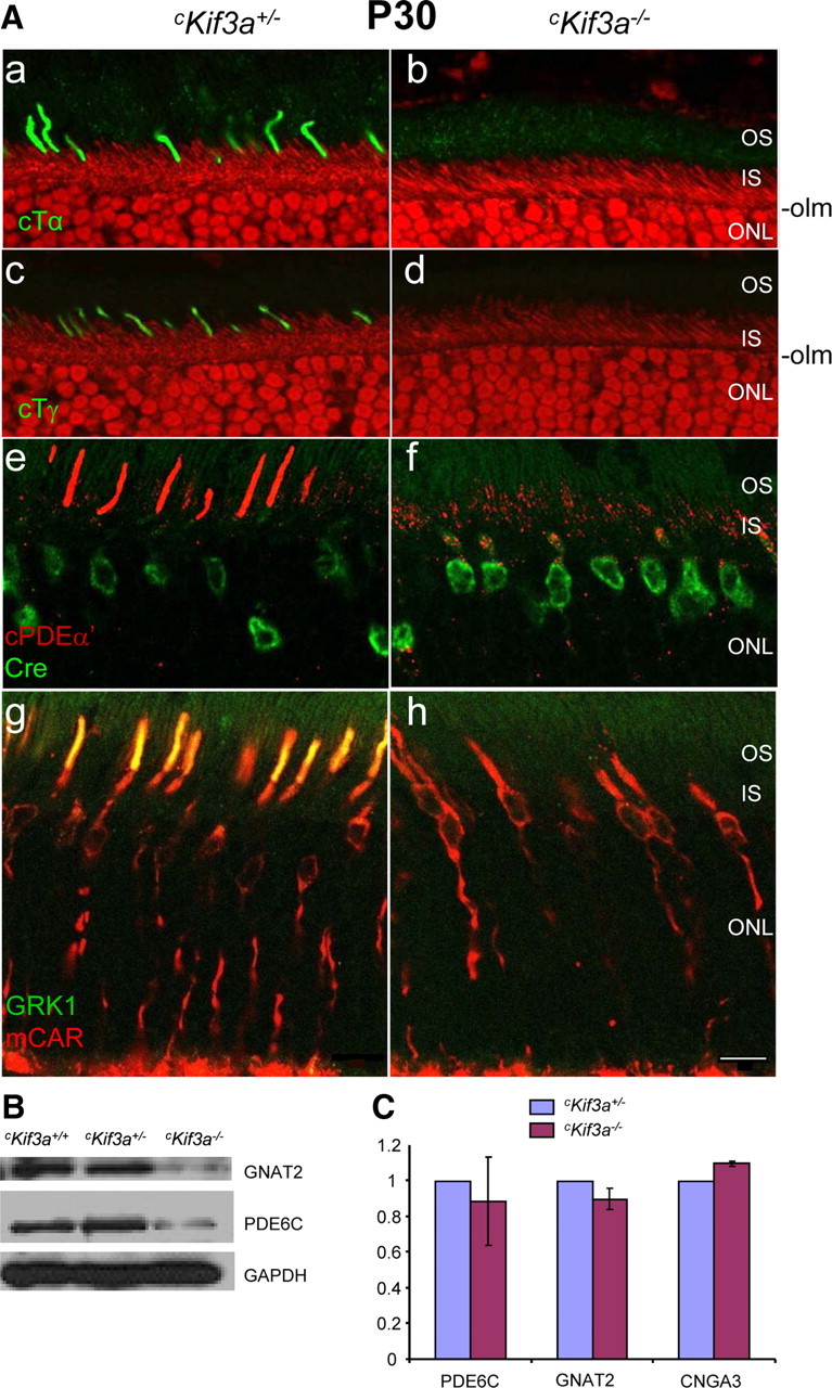
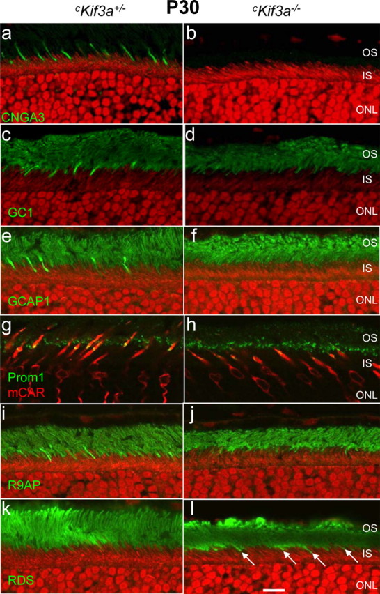
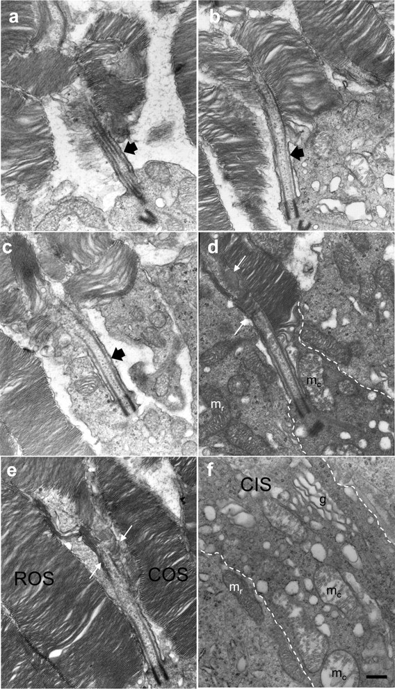
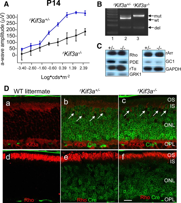
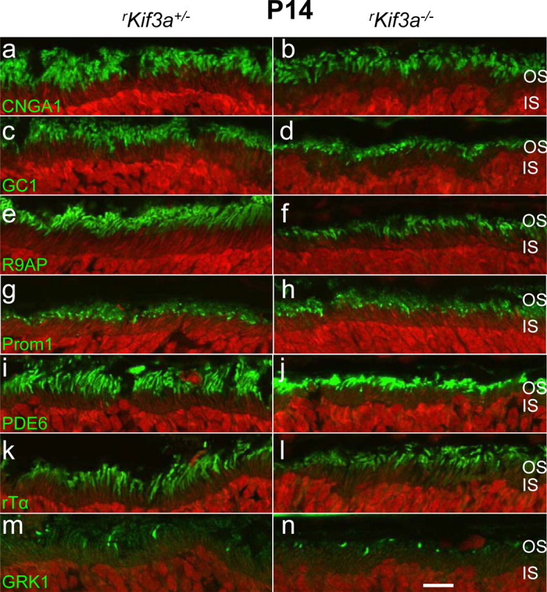
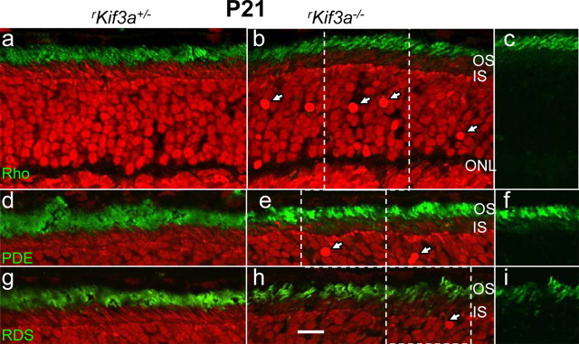
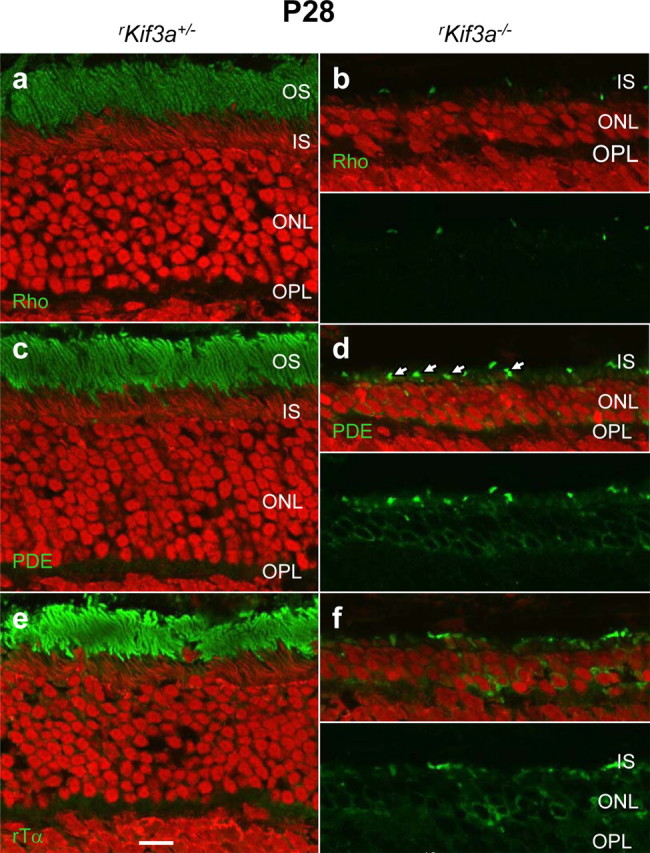
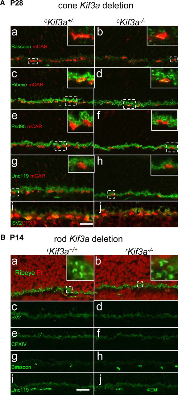
Heterotrimeric kinesin-II is a molecular motor localized to the inner segment, connecting cilium and axoneme of mammalian photoreceptors. Our purpose was to identify the role of kinesin-II in anterograde intraflagellar transport by photoreceptor-specific deletions of kinesin family member 3A (KIF3A), its obligatory motor subunit. In cones lacking KIF3A, membrane proteins involved in phototransduction did not traffic to the outer segments resulting in complete absence of a photopic electroretinogram and progressive cone degeneration. Rod photoreceptors lacking KIF3A degenerated rapidly between 2 and 4 weeks postnatally, but the phototransduction components including rhodopsin trafficked to the outer segments during the course of degeneration. Furthermore, KIF3A deletion did not affect synaptic anterograde trafficking. The results indicate that trafficking of membrane proteins to the outer segment is dependent on kinesin-II in cone, but not rod photoreceptors, even though rods and cones share similar structures, and closely related phototransduction polypeptides.


Figures

Figure 1.
Figure 1.
Generation and phenotypes of cKif3a+/− and cKif3a−/− mice. A, Generation of cone-specific Kif3a knock-outs (cKif3a−/−) by mating of Kif3aflox/flox mice with transgenic mice expressing Cre recombinase under the control of the human red/green opsin (Hrgp) promoter. a, Diagram of exon 1 and exon 2 of the Kif3a gene; b, the floxed gene; c, the targeted gene with a deletion of exon 2. Primers 1–3 are used for genotyping the Kif3a alleles (Marszalek et al., 2000). B, Photopic ERG at P30. Black trace, The cKif3a+/− response; red trace, the cKif3a−/− response to a single flash (intensity, 25 db or 2.9 log cd s m−2). C, Confocal immunolocalization of ML-opsin and S-opsin in cone photoreceptors of 1-month-old cKif3a+/− (a, c) and cKif3a−/− mice (b, d). In a–d, sections were probed with anti-Cre antibody (green); in a and b, sections were probed with anti-ML-opsin (red), and in c and d, with anti S-opsin (red). Note that Cre recombinase is expressed in cone nuclei exclusively. ML-opsin and S-opsin are mislocalized to the inner segments, axons, and synaptic pedicles (white arrows) of cKif3a−/− cones. Scale bar, 10 μm. IS, Inner segment; OPL, outer plexiform layer.
Figure 2.
Figure 2.
Immunolocalization of cone Tα, Tγ, PDE6α′, GRK1, and mCar in cKif3a+/− and cKif3a−/− cones. A, Frozen sections were probed with either anti-cone Tα (green; a, b), anti-cone Tγ (green; c, d), anti-cone PDE6α′ (red; e, f), or anti-GRK1 (green; g, h) antibodies. Some sections were contrasted with propidium iodide (red) to define the outer limiting membrane (olm) and the inner segments (a–d). Note the normal distributions of cone Tα, cone Tγ, and cone PDE6α′ in cKif3a+/− COS, but absence (b, d, h) or mislocalization to the inner segments (f) of cKif3a−/− cones. In contrast, sections (g, h) probed simultaneously with antibodies directed against cone arrestin (mCAR; red) and anti-GRK1 (green) revealed that cone arrestin distributes freely in cKif3a+/− and cKif3a−/− cones. Cone arrestin and GRK1 colocalize (yellow; g) in OS of cKif3a+/−, but not cKif3a−/− cones. IS, Inner segments. Scale bars, 10 μm. B, Immunoblots of cKif3a+/−, and cKif3a−/− retina lysates probed with anti-cone Tα, and anti-cone PDE6α′ antibodies. In each immunoblot, GAPDH was included as an internal control. Note cone transducin and PDE6α′ subunit levels are reduced in cKif3a−/−. C, Semiquantitative real-time RT/PCR of cone Pde6a (PDE6α′), Gnat2 (cone Tα), and Cnga3 mRNA of cKif3a+/− and cKif3a−/− retinas. Each sample was determined in duplicate from two retinas of different animals of the same genotype. PCRs were run in parallel with Gapdh-specific primers as a standard. Note that mRNA levels are unaffected. Scale bar, 10 μm.
Figure 3.
Figure 3.
Distribution of outer segment membrane proteins in P30 Kif3a-deleted cones. Sections of cKif3a+/− (a, c, e, g, i, k) and cKif3a−/− retinas (b, d, f, h, j, l) were probed with antibodies recognizing CNGA3 (a, b), GC1 (c, d), GCAP1 (e, f), prominin-1 (g, h), R9AP (i, j), and peripherin/rds (k, l). Antigen localizations are visualized using FITC-conjugated secondary antibody (green). The ONL of some sections was contrasted with propidium iodide (a–f, i–l), whereas in other sections, cone cells were identified with anti-cone arrestin (red; g, h). Note that, in cKif3a−/− cones, membrane proteins were severely reduced or completely absent, with the exception of peripherin/rds (l, arrows). Scale bar, 10 μm.
Figure 4.
Figure 4.
Fine structure of Kif3a-deleted cones at postnatal day 13. Proximal COSs reveal that connecting cilia (black arrows; a–c) develop, although outer segment membranes appear misaligned. Misalignment was most severe in cones inferior to the optic nerve, and less severe in cones located in the superior midperiphery. Examples of COSs found in various stages of membrane disorganization: a, immediately inferior to the optic nerve and advanced; b, c, intermediate; and d, e, early in degeneration. The plasma membrane (dashed white line; d, f) defines the extent of the distal cone inner segment (CIS), in which cone mitochondria (mc) with disrupted cristae were distinct from rod mitochondria (mr). A cascade of five to seven overlapping membrane discs appears at the expanded axoneme (between pair of white arrows; d, e). More proximally, in a cKif3a−/− cone inner segment (f, between dashed white lines), vesicles of heterogeneous diameter were dispersed among mitochondria (mc) and a Golgi apparatus. Scale bar, 0.2 μm (for a–f).
Figure 5.
Figure 5.
KIF3A deletion in P14 rod photoreceptors. A, Scotopic (dark-adapted) electroretinogram of P14 rKif3a+/− and rKif3a−/− mice. The rKif3a−/− a-wave amplitude, indicative of rod function, is attenuated relative to the WT a-wave response at all intensities tested. Error bars indicate SEM. B, PCR-based genotyping of rKif3a+/− (lane 2) and rKif3a−/− mice (lane 3) with primers P1, P2, and P3 and retina DNA as a template (see Materials and Methods) (Fig. 1A). P1 and P2 amplify the WT fragment (wt) and the mutant (insertion of loxP) fragment (mut) from rKif3a+/− DNA, but only the mutant fragment from rKif3a−/− DNA. P1 and P3 amplify the deletion fragment (del) appearing after excision of exon 2. Both the heterozygous and homozygous floxed mice show the deletion fragment at P14. Lane 1, MW standards. C, Immunoblots. Polypeptides of rKif3a+/− and rKif3a−/− retina lysates were separated by SDS-PAGE, blotted, and probed with anti-rhodopsin, anti-rod Tα, anti-rod PDE6, anti-GRK1, anti-rod arrestin, and anti-GC1. Anti-GAPDH served as a loading control. Note that the proteins levels are very similar in both from rKif3a+/− (+/−) and rKif3a−/− (−/−) lanes. D, Confocal immunolocalization of KIF3A and rhodopsin in P14 littermate WT (a, d), rKif3a+/− (b, e), and rKif3a−/− (c, f) cryosections imaged in the midperiphery. Retina sections were probed simultaneously with anti-Cre recombinase monoclonal antibody (green; b, c, e, f) and either anti-KIF3A (red; a–c) or anti-rhodopsin (red; d–f) polyclonal antibody. The white arrows (b, c) indicate examples of cone nuclei that do not express Cre recombinase. KIF3A is essentially undetectable in rKif3a−/− rod inner segments but present in cone inner segments, which are unaffected by the rod-specific deletion. Note prominent immunolabel for rhodopsin over rKif3a−/− rod outer segments that are reduced in length relative to WT. Several pyknotic nuclei were observed in the proximal rKif3a−/− ONL. Scale bar: a–f, 10 μm.
Figure 6.
Figure 6.
Confocal localization of phototransduction proteins. Sections of rKif3a+/− (a, c, e, g, i, k, m) and rKif3a−/− (b, d, f, h, j, l, n) retina were probed with anti-CNGA1 (a, b), anti-GC1 (c, d), anti-R9AP (e, f), anti-prom1 (g, h), anti-rod PDE6 (i, j), anti-rod Tα (k, l), and anti-GRK1 (m, n) antibodies. All panels show normal transport and localization of membrane proteins to the outer segments. Scale bar, 10 μm.
Figure 7.
Figure 7.
Trafficking of rhodopsin, PDE6 subunits, and peripherin/rds in P21 rKif3a−/− rods. Left column (a, d, g), rKif3a+/− retina sections; middle column (b, e, h), rKif3a−/− retina sections; and right column (c, f, i), portions of corresponding sections (b, e, h), green channel only. Sections a–c, Anti-rhodopsin; sections d–f, anti-PDE6 antibody; sections g–i, anti-peripherin/rds antibody. Note that rhodopsin traffics normally and does not mislocalize at P21. The white arrows indicate pyknotic nuclei. Scale bar, 10 μm.
Figure 8.
Figure 8.
Rapid degeneration of rKif3a−/− rod photoreceptors at P28. Sections of rKif3a+/− (a, c, e) and rKif3a−/− (b, d, f) retina were probed with anti-rhodopsin (a, b), anti-rod PDE6 (c, d), and anti-transducin α-subunit antibodies (e, f). Rod outer segments in rKif3a−/− sections were absent and the ONL is reduced to three to four rows. The bottom panels in b, d, and f show green channel only. In b, very little rhodopsin remains in the outer segments. In d, punctuate green fluorescence (arrows) indicates remnants of cone outer segments (labeled with MOE, an antibody that recognizes both rod and cone PDE6). Scale bar, 10 μm.
Figure 9.
Figure 9.
Synaptic terminal membrane proteins appear unaffected by KIF3A deletion. A, KIF3A deletion in cones. cKif3a+/− (a, c, e, g, i) and cKif3a−/− (b, d, f, h, j) sections were probed with anti-bassoon (green) and anti-mCAR (red) antibodies (a, b), anti-ribeye (green) and anti mCAR (c, d), anti-PSD95 (green) and anti-mCAR (e, f), anti-Unc119 (green) and anti-mCAR antibodies (g, h), and with anti-SV2 (green) and anti-CPX-III (red) antibodies. The insets show enlargements identified by broken lines. B, KIF3A deletion in rods. rKif3a+/− (a, c, e, g, i) and rKif3a−/− (b, d, f, h, j) sections were probed with ribeye antibody (a, b), anti-SV2 antibody (c, d), anti-CPX-IV (e, f), anti-bassoon (g, h), and anti-Unc119 antibodies (i, j). Nuclei of the ONL and INL were counterstained with propidium iodide to clearly mark the OPL region (a, b). The insets (a, b) show enlarged regions identified by broken lines. Scale bar, 10 μm.

References

    1. Abd-El-Barr MM, Sykoudis K, Andrabi S, Eichers ER, Pennesi ME, Tan PL, Wilson JH, Katsanis N, Lupski JR, Wu SM. Impaired photoreceptor protein transport and synaptic transmission in a mouse model of Bardet-Biedl syndrome. Vision Res. 2007;47:3394–3407. - PMC - PubMed
    1. Anderson DH, Fisher SK, Steinberg RH. Mammalian cones: disc shedding, phagocytosis, and renewal. Invest Ophthalmol Vis Sci. 1978;17:117–133. - PubMed
    1. Applebury ML, Antoch MP, Baxter LC, Chun LL, Falk JD, Farhangfar F, Kage K, Krzystolik MG, Lyass LA, Robbins JT. The murine cone photoreceptor: a single cone type expresses both S and M opsins with retinal spatial patterning. Neuron. 2000;27:513–523. - PubMed
    1. Baehr W, Karan S, Maeda T, Luo DG, Li S, Bronson JD, Watt CB, Yau KW, Frederick JM, Palczewski K. The function of guanylate cyclase 1 and guanylate cyclase 2 in rod and cone photoreceptors. J Biol Chem. 2007;282:8837–8847. - PMC - PubMed
    1. Baker SA, Freeman K, Luby-Phelps K, Pazour GJ, Besharse JC. IFT20 links kinesin II with a mammalian intraflagellar transport complex that is conserved in motile flagella and sensory cilia. J Biol Chem. 2003;278:34211–34218. - PubMed

Publication types

MeSH terms

LinkOut - more resources