. 2009 Nov 11;29(45):14334-41.
doi: 10.1523/JNEUROSCI.2770-09.2009.

Neuroprotective and axon growth-promoting effects following inflammatory stimulation on mature retinal ganglion cells in mice depend on ciliary neurotrophic factor and leukemia inhibitory factor

Affiliations

Neuroprotective and axon growth-promoting effects following inflammatory stimulation on mature retinal ganglion cells in mice depend on ciliary neurotrophic factor and leukemia inhibitory factor

Marco Leibinger et al. J Neurosci. .

Abstract

After optic nerve injury retinal ganglion cells (RGCs) normally fail to regenerate axons in the optic nerve and undergo apoptosis. However, lens injury (LI) or intravitreal application of zymosan switch RGCs into an active regenerative state, enabling these neurons to survive axotomy and to regenerate axons into the injured optic nerve. Several factors have been proposed to mediate the beneficial effects of LI. Here, we investigated the contribution of glial-derived ciliary neurotrophic factor (CNTF) to LI-mediated regeneration and neuroprotection using wild-type and CNTF-deficient mice. In wild-type mice, CNTF expression was strongly upregulated in retinal astrocytes, the JAK/STAT3 pathway was activated in RGCs, and RGCs were transformed into an active regenerative state after LI. Interestingly, retinal LIF expression was correlated with CNTF expression after LI. In CNTF-deficient mice, the neuroprotective and axon growth-promoting effects of LI were significantly reduced compared with wild-type animals, despite an observed compensatory upregulation of LIF expression in CNTF-deficient mice. The positive effects of LI and also zymosan were completely abolished in CNTF/LIF double knock-out mice, whereas LI-induced glial and macrophage activation was not compromised. In culture CNTF and LIF markedly stimulated neurite outgrowth of mature RGCs. These data confirm a key role for CNTF in directly mediating the neuroprotective and axon regenerative effects of inflammatory stimulation in the eye and identify LIF as an additional contributing factor.


Figures

Figure 1.
Figure 1.
Changes in the expression of CNTF and LIF, as well as JAK/STAT3 pathway activation, in the murine retina after ONC and LI. A, Immunohistochemical staining of an untreated control retina (con), a retina 5 d after optic nerve crush (onc), and onc + lens injury (li) from wild-type mice as indicated using an anti-CNTF (red) and an anti-GFAP (green) antibody. Scale bar, 50 μm. GCL, ganglion cell layer; INL, inner nuclear layer. B, Western blot analysis of retinal lysates from wild-type (wt) and CNTF-deficient (cntf−/−) mice 5 d after onc, onc + li, or no previous treatment (con) using specific antibodies against GFAP, GAP43, LIF, phospho-STAT3 (pSTAT3), or CNTF. Tubulin served as a loading control. C, Immunohistochemical staining of astrocytes of an untreated control retina, a retina 5 d after onc, and after onc + li of wt and cntf−/− mice using an anti-LIF (R&D) (red) and anti-GFAP antibody (green). Scale bar, 25 μm. D, Quantitative real-time PCR. LIF expression levels were quantified relative to GAPDH in wt and cntf−/− mice 5 d after onc, onc + li, or untreated animals (−). *p < 0.05; ***p < 0.001. E, Western blot analysis of retinal lysates from CNTF/LIF double knock-out (cntf−/−/lif−/−) mice 5 d after onc, onc + li, or no previous treatment (−) using specific antibodies against GAP43, GFAP, and pSTAT3. Tubulin expression verified that the same amount of retinal protein was loaded per lane.
Figure 2.
Figure 2.
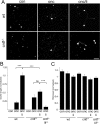
Regenerative state of RGCs 5 d after ONC and ONC + LI in mature wild-type and CNTF-deficient mice. A, Dissociated retinal cell cultures immunostained with an antibody against βIII-tubulin, showing spontaneously regenerating RGCs after 24 h in culture. Wild-type (wt) and CNTF-deficient (cntf−/−) mice had received ONC (onc), ONC + LI (onc + li), or no treatment (con) 5 d previously. Scale bar, 50 μm. B, Quantitation of neurite outgrowth of groups in A and of CNTF/LIF double knock-out (cntf−/−/lif−/−) mice 5 d after ONC + LI, indicated as average neurite length per RGC. C, Quantitation of RGCs per well after 24 h in culture, demonstrating no significant differences between groups. Significance between groups: ***p < 0.001; ns., nonsignificant.
Figure 3.
Figure 3.
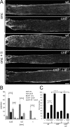
Axon regeneration and survival of RGCs in vivo after LI in wild-type, CNTF-deficient and CNTF/LIF double knock-out mice. A, Longitudinal sections through the optic nerve showing GAP43-positive axons distal to the injury site (asterisk) in wild-type (wt), CNTF-deficient (cntf −/−) and CNTF/LIF double knock-out (cntf −/−/lif −/−) mice 2 weeks after ONC alone (onc) or ONC + LI (onc + li). Scale bar, 100 μm. B, Quantification of axon regeneration (number of axons growing 0.25, 0.5, and 1 mm beyond the injury site per optic nerve) 2 weeks after ONC alone or ONC + LI. C, Quantification of surviving RGCs (βIII-tubulin-positive RGCs per retinal cross-section) 2 weeks after ONC alone, ONC + LI, or untreated retinas (−). *p < 0.05; *** p < 0.001.
Figure 4.
Figure 4.
Neurite growth-promoting effects of LIF and CNTF on mature RGCs in culture. A, Quantitation of neurite outgrowth of RGCs in the presence of increasing concentrations of LIF (as indicated), CNTF and an anti-LIF-antibody (α-LIF) for 3 d. Values were normalized to untreated controls. Treatment effects compared with the group treated with vehicle only. *p < 0.05; ***p < 0.001. B, Quantitation of RGCs per well after 3 d in culture, demonstrating no significant differences between groups.

Similar articles

Cited by

References

    1. Banner LR, Moayeri NN, Patterson PH. Leukemia inhibitory factor is expressed in astrocytes following cortical brain injury. Exp Neurol. 1997;147:1–9. - PubMed
    1. Benowitz L, Yin Y. Rewiring the injured CNS: lessons from the optic nerve. Exp Neurol. 2008;209:389–398. - PMC - PubMed
    1. Benowitz LI, Yin Y. Combinatorial treatments for promoting axon regeneration in the CNS: strategies for overcoming inhibitory signals and activating neurons' intrinsic growth state. Dev Neurobiol. 2007;67:1148–1165. - PubMed
    1. Berkelaar M, Clarke DB, Wang YC, Bray GM, Aguayo AJ. Axotomy results in delayed death and apoptosis of retinal ganglion cells in adult rats. J Neurosci. 1994;14:4368–4374. - PMC - PubMed
    1. Berry M, Carlile J, Hunter A. Peripheral nerve explants grafted into the vitreous body of the eye promote the regeneration of retinal ganglion cell axons severed in the optic nerve. J Neurocytol. 1996;25:147–170. - PubMed

Publication types

MeSH terms

LinkOut - more resources