

Differences in the TGF-{beta}1-induced profibrotic response of anterior and posterior corneal keratocytes in vitro
- PMID: 19907023
- PMCID: PMC2846206
- DOI: 10.1167/iovs.09-3823
Differences in the TGF-{beta}1-induced profibrotic response of anterior and posterior corneal keratocytes in vitro
Abstract
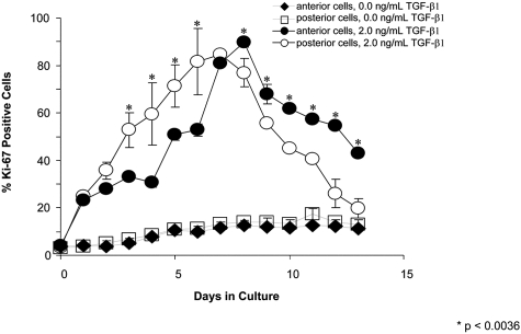
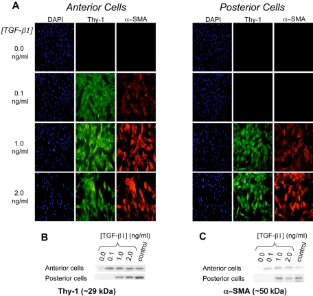
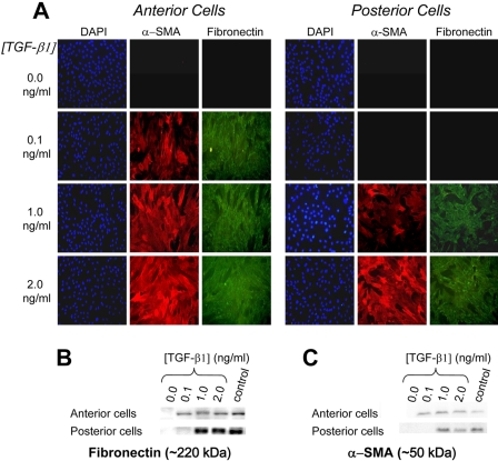
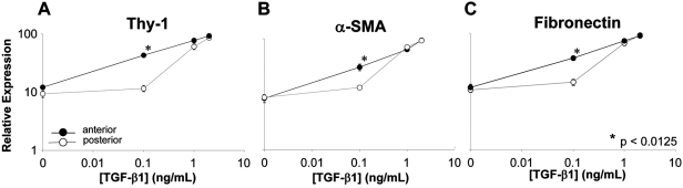
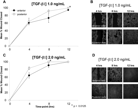
Purpose. To characterize phenotypic differences between anterior and posterior corneal keratocytes after stimulation with the profibrotic agent transforming growth factor-beta1 (TGF-beta1) in vitro. Methods. Sixteen corneas from healthy felines were obtained immediately after death. Lamellar dissection was performed to separate the anterior and posterior stroma at approximately 50% depth either manually (n = 2) or with a Moria microkeratome (300-mum head; n = 14). Cells from the anterior and posterior stroma were cultured separately but under identical conditions. Using immunohistochemistry and Western blot techniques, Ki-67 staining and relative expression of Thy-1, alpha smooth muscle actin (alpha-SMA), and fibronectin were assessed after stimulation with different TGF-beta1 concentrations. In addition, anterior and posterior cells cultured in different concentrations of TGF-beta1 were wounded with a razor blade, and the wound area and time to closure were determined. Results. Stimulation by all concentrations of TGF-beta1 increased the proportion of Ki-67-positive cells in anterior and posterior cell cultures, but this increase was noted earlier in posterior cells than in anterior cells. Increasing TGF-beta1 concentration also increased the relative expression of Thy-1, alpha-SMA, and fibronectin in anterior and posterior fibroblasts. However, anterior cells expressed these fibrotic markers at lower TGF-beta1 concentrations than did posterior keratocytes. After mechanical wounding, posterior cells closed the wound area faster than did anterior cells at all concentrations of TGF-beta1. Conclusions. The present experiments show that anterior and posterior corneal keratocytes exhibit different sensitivities to the profibrotic growth factor TGF-beta1. This heterogeneity of keratocyte response may impact wound closure after mechanical wounding.
Figures
Similar articles
-
Responses of cultured human keratocytes and myofibroblasts to ethyl pyruvate: a microarray analysis of gene expression.Invest Ophthalmol Vis Sci. 2010 Jun;51(6):2917-27. doi: 10.1167/iovs.09-4498. Epub 2010 Jan 6. Invest Ophthalmol Vis Sci. 2010. PMID: 20053976 Free PMC article.
-
IL-1 and TGF-β Modulation of Epithelial Basement Membrane Components Perlecan and Nidogen Production by Corneal Stromal Cells.Invest Ophthalmol Vis Sci. 2018 Nov 1;59(13):5589-5598. doi: 10.1167/iovs.18-25202. Invest Ophthalmol Vis Sci. 2018. PMID: 30480706 Free PMC article.
-
IGF-II is present in bovine corneal stroma and activates keratocytes to proliferate in vitro.Exp Eye Res. 2008 Mar;86(3):506-11. doi: 10.1016/j.exer.2007.12.004. Epub 2007 Dec 23. Exp Eye Res. 2008. PMID: 18237730 Free PMC article.
-
Transforming growth factor beta-3 localization in the corneal response to epithelial-stromal injury and effects on corneal fibroblast transition to myofibroblasts.Exp Eye Res. 2023 Oct;235:109631. doi: 10.1016/j.exer.2023.109631. Epub 2023 Aug 25. Exp Eye Res. 2023. PMID: 37633325
-
The effect of growth factor supplementation on corneal stromal cell phenotype in vitro using a serum-free media.Exp Eye Res. 2016 Oct;151:26-37. doi: 10.1016/j.exer.2016.07.015. Epub 2016 Jul 22. Exp Eye Res. 2016. PMID: 27456135
Cited by
-
More than Meets the Eye: The Aryl Hydrocarbon Receptor is an Environmental Sensor, Physiological Regulator and a Therapeutic Target in Ocular Disease.Front Toxicol. 2022 Mar 3;4:791082. doi: 10.3389/ftox.2022.791082. eCollection 2022. Front Toxicol. 2022. PMID: 35295218 Free PMC article. Review.
-
Ocular fibroblast diversity: implications for inflammation and ocular wound healing.Invest Ophthalmol Vis Sci. 2011 Jul 1;52(7):4859-65. doi: 10.1167/iovs.10-7066. Invest Ophthalmol Vis Sci. 2011. PMID: 21571679 Free PMC article.
-
Fibroblastic and bone marrow-derived cellularity in the corneal stroma.Exp Eye Res. 2021 Jan;202:108303. doi: 10.1016/j.exer.2020.108303. Epub 2020 Oct 14. Exp Eye Res. 2021. PMID: 33068626 Free PMC article.
-
Matrix glycosaminoglycans and proteoglycans in human cornea organoids and similarities with fetal corneal stages.Ocul Surf. 2025 Jan;35:68-80. doi: 10.1016/j.jtos.2024.11.007. Epub 2024 Nov 28. Ocul Surf. 2025. PMID: 39615587
-
Moxifloxacin modifies corneal fibroblast-to-myofibroblast differentiation.Br J Pharmacol. 2013 Mar;168(6):1341-54. doi: 10.1111/bph.12015. Br J Pharmacol. 2013. PMID: 23072440 Free PMC article.
References
-
- Watsky M. Keratocyte gap junctional communication in normal and wounded rabbit corneas and human corneas. Invest Ophthalmol Vis Sci 1995;36:2568–2576 - PubMed
-
- Snyder MC, Bergmanson JP, Doughty MJ. Keratocytes: no more the quiet cells. J Am Optom Assn 1998;69:180–187 - PubMed
-
- Brown D, Chwa M, Escobar M, Kenney MC. Characterization of the major matrix degrading metalloproteinase of human corneal stroma: evidence for an enzyme/inhibitor complex. Exp Eye Res 1991;52:5–16 - PubMed
-
- Pei Y, Sherry DM, McDermott AM. Thy-1 distinguished human corneal fibroblasts and myofibroblasts from keratocytes. Exp Eye Res 2004;79:705–712 - PubMed
Publication types
MeSH terms
Substances
Grants and funding
- K23 EY019353/EY/NEI NIH HHS/United States
- EY017123/EY/NEI NIH HHS/United States
- P30 ES001247/ES/NIEHS NIH HHS/United States
- HL75432/HL/NHLBI NIH HHS/United States
- P30 EY001319/EY/NEI NIH HHS/United States
- R03 HL095402/HL/NHLBI NIH HHS/United States
- HL095402/HL/NHLBI NIH HHS/United States
- T32 HL066988/HL/NHLBI NIH HHS/United States
- R01 EY015836/EY/NEI NIH HHS/United States
- R01 EY017123/EY/NEI NIH HHS/United States
- P0EY01319F/EY/NEI NIH HHS/United States
- ES01247/ES/NIEHS NIH HHS/United States
- L30 EY019138/EY/NEI NIH HHS/United States
- R01 HL075432/HL/NHLBI NIH HHS/United States
- EY015836/EY/NEI NIH HHS/United States
- KL2 RR024136/RR/NCRR NIH HHS/United States
LinkOut - more resources
Full Text Sources
Miscellaneous
