. 2010 Jan 8;285(2):1569-76.
doi: 10.1074/jbc.M109.060327. Epub 2009 Nov 11.

Mode of binding of the tuberculosis prodrug isoniazid to heme peroxidases: binding studies and crystal structure of bovine lactoperoxidase with isoniazid at 2.7 A resolution

Affiliations

Mode of binding of the tuberculosis prodrug isoniazid to heme peroxidases: binding studies and crystal structure of bovine lactoperoxidase with isoniazid at 2.7 A resolution

Amit K Singh et al. J Biol Chem. .

Abstract

Isoniazid (INH) is an anti-tuberculosis prodrug that is activated by mammalian lactoperoxidase and Mycobacterium tuberculosis catalase peroxidase (MtCP). We report here binding studies, an enzyme assay involving INH, and the crystal structure of the complex of bovine lactoperoxidase (LPO) with INH to illuminate binding properties and INH activation as well as the mode of diffusion and interactions together with a detailed structural and functional comparison with MtCP. The structure determination shows that isoniazid binds to LPO at the substrate binding site on the distal heme side. The substrate binding site is connected to the protein surface through a long hydrophobic channel. The acyl hydrazide moiety of isoniazid interacts with Phe(422) O, Gln(423) O(epsilon1), and Phe(254) O. In this arrangement, pyridinyl nitrogen forms a hydrogen bond with a water molecule, W-1, which in turn forms three hydrogen bonds with Fe(3+), His(109) N(epsilon2), and Gln(105) N(epsilon2). The remaining two sides of isoniazid form hydrophobic interactions with the atoms of heme pyrrole ring A, C(beta) and C(gamma) atoms of Glu(258), and C(gamma) and C(delta) atoms of Arg(255). The binding studies indicate that INH binds to LPO with a value of 0.9 x 10(-6) m for the dissociation constant. The nitro blue tetrazolium reduction assay shows that INH is activated by the reaction of LPO-H(2)O(2) with INH. This suggests that LPO can be used for INH activation. It also indicates that the conversion of INH into isonicotinoyl radical by LPO may be the cause of INH toxicity.


Figures

FIGURE 1.
FIGURE 1.
Stereoview of the observed |FoFc| difference Fourier density (countered at 2.5σ) for the INH molecule prior to inclusion of INH in the model. This figure was drawn using PyMol (49).
FIGURE 2.
FIGURE 2.
Plot showing INH binding to LPO. The different concentrations (1–20 μm) of INH were incubated with 5 μm LPO for 12 h to make sure that the equilibrium was achieved. The binding of INH was detected by taking absorbance of LPO (heme) at 412 nm. The data were fitted using SigmaPlot version 8.0 (20). The absorbance of LPO (heme) without INH (A0) and with INH (A) were measured. The dissociation constant (Kd) of LPO with INH was determined from the non-linear fit curve.
FIGURE 3.
FIGURE 3.
NBT reduction by the product of the reaction LPO-H2O2 with INH. The reaction mixture consisted of 0.2 mm NBT, 33 μg of LPO, and 100 μm H2O2 in 3.0 ml of 0.05 m phosphate buffer (pH 7.0) at 298 K. The reaction was initiated by adding 9.6 mm INH. The change A560 with time was recorded. The reference cuvettes contained the same concentrations of NBT, LPO, and INH.
FIGURE 4.
FIGURE 4.
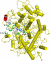
The overall folding of the protein shown as a schematic diagram, where α-helices are indicated as cylinders and numbered. Helix H2a (red) is a unique element of the structure of LPO. The two anti-parallel β-strands are drawn as arrows. Heme iron (Fe) is shown in blue. Residues involved in the hydrogen bonding with INH are also shown. Two water molecules, W-1 and W-89, are indicated as part of the hydrogen-bonded network. An important salt bridge between Glu130 and His426 that works as an anchor for the loop 421–430 is also shown. The dotted lines indicate hydrogen bonds.
FIGURE 5.
FIGURE 5.
The position of six water molecules, W-1, W-2′, W-3′, W-4′, W-5′, and W-6′ in the substrate binding site on the distal side. The water molecules W-2′, W-4′, W-5′, and W-6′, indicated by hollow red circles, are replaced by INH, whereas W-1 and W-3′ remain and participate in the hydrogen-bonded network at the binding site.
FIGURE 6.
FIGURE 6.
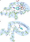
Schematic drawing showing the hydrophobic channels in heme peroxidases. a, substrate binding site on the distal heme side in LPO with bound INH molecule. The hydrophobic channel with arrows indicating the passage for the ligands is also shown. b, substrate binding site in MtCP and the hydrophobic channel. The INH position is superimposed from the complex of LPO with INH. The side chain of Ser315 is rotated to form the hydrogen bond with INH. c, substrate binding site in CcP on the distal side with a bound INH molecule. The substrate binding site is located very close to the protein surface.
FIGURE 6.
FIGURE 6.
Schematic drawing showing the hydrophobic channels in heme peroxidases. a, substrate binding site on the distal heme side in LPO with bound INH molecule. The hydrophobic channel with arrows indicating the passage for the ligands is also shown. b, substrate binding site in MtCP and the hydrophobic channel. The INH position is superimposed from the complex of LPO with INH. The side chain of Ser315 is rotated to form the hydrogen bond with INH. c, substrate binding site in CcP on the distal side with a bound INH molecule. The substrate binding site is located very close to the protein surface.

Similar articles

Cited by

References

    1. Kimura S., Ikeda-Saito M. (1988) Proteins Struct. Funct. Genet. 3, 113–120 - PubMed
    1. Welinder K. G. (1992) Curr. Opin. Struct. Biol. 2, 388–393
    1. Zeng J., Fenna R. E. (1992) J. Mol. Biol. 226, 185–207 - PubMed
    1. Singh A. K., Singh N., Sharma S., Singh S. B., Kaur P., Bhushan A., Srinivasan A., Singh T. P. (2008) J. Mol. Biol. 376, 1060–1075 - PubMed
    1. Gajhede M., Schuller D. J., Henriksen A., Smith A. T., Poulos T. L. (1997) Nat. Struct. Biol. 4, 1032–1038 - PubMed

Publication types

LinkOut - more resources