. 2009 Dec;119(12):3797-806.
doi: 10.1172/JCI37976. Epub 2009 Nov 9.

PKC inhibition ameliorates the cardiac phenotype in a mouse model of myotonic dystrophy type 1

Affiliations

PKC inhibition ameliorates the cardiac phenotype in a mouse model of myotonic dystrophy type 1

Guey-Shin Wang et al. J Clin Invest. 2009 Dec.

Abstract

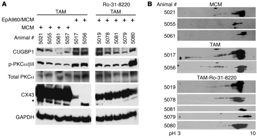
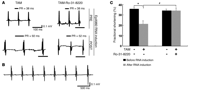
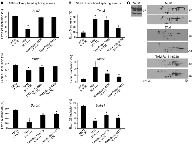
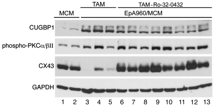
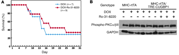
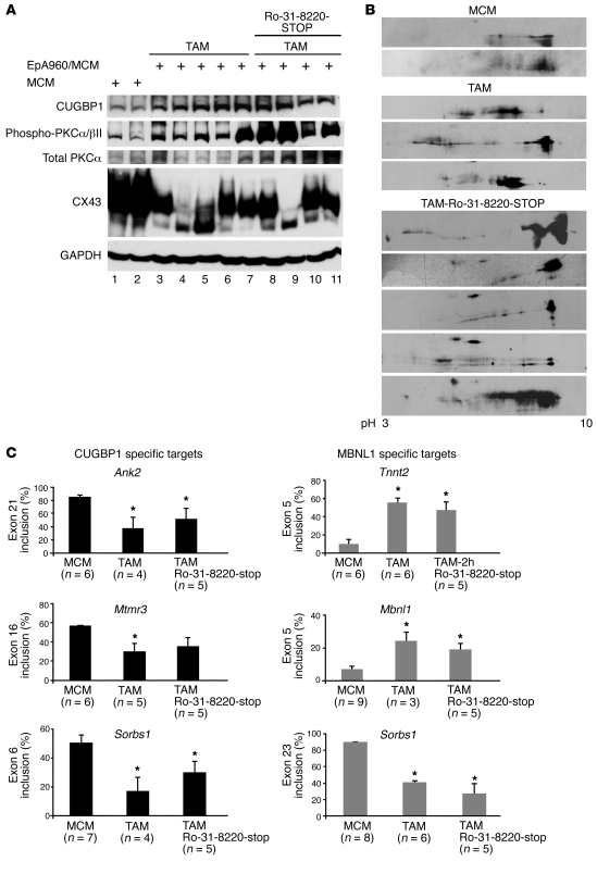
Cardiac complications are a common cause of death in individuals with the inherited multisystemic disease myotonic dystrophy type 1 (DM1). A characteristic molecular feature of DM1 is misregulated alternative splicing due to disrupted functioning of the splicing regulators muscleblind-like 1 (MBNL1) and CUG-binding protein 1 (CUGBP1). CUGBP1 is upregulated in DM1 due to PKC pathway activation and subsequent CUGBP1 protein hyperphosphorylation and stabilization. Here, we blocked PKC activity in a heart-specific DM1 mouse model to determine its pathogenic role in DM1. Animals given PKC inhibitors exhibited substantially increased survival that correlated with reduced phosphorylation and decreased steady-state levels of CUGBP1. Functional studies demonstrated that PKC inhibition ameliorated the cardiac conduction defects and contraction abnormalities found in this mouse model. The inhibitor also reduced misregulation of splicing events regulated by CUGBP1 but not those regulated by MBNL1, suggesting distinct roles for these proteins in DM1 cardiac pathogenesis. The PKC inhibitor did not reduce mortality in transgenic mice with heart-specific CUGBP1 upregulation, indicating that PKC inhibition did not have a general protective effect on PKC-independent CUGBP1 increase. Our results suggest that pharmacological blockade of PKC activity mitigates the DM1 cardiac phenotype and provide strong evidence for a role for the PKC pathway in DM1 pathogenesis.


Figures

Figure 1
Figure 1. PKC inhibition improves the mortality rate of mice expressing EpA960(R) RNA.
(A) Kaplan-Meier survival curve on animals receiving the PKC inhibitor, Ro-31-8220. Percentage survival of tamoxifen-induced EpA960/MCM mice treated with Ro-31-8220 (EpA960/MCM, TAM-Ro-31-8220) or mock injected using saline (EpA960/MCM, TAM), compared with control mice, expressing only Cre treated with tamoxifen and Ro-31-8220 (MCM, TAM-Ro-31-8220). (B) Expression of EpA960(R) RNA after Cre-mediated recombination in TAM-induced mice given Ro-31-8220 (TAM-Ro-31-8220) or saline (TAM) was analyzed by quantitative RT-PCR using TaqMan (mean ± SEM). (C) HW/BW ratio of mice from each group (mean ± SEM). *P < 0.05 versus MCM control.
Figure 2
Figure 2. Effect of Ro-31-8220 administration on PKC activation and CUGBP1 and connexin 43 expression.
(A) Western blots of CUGBP1 (50 kDa), phospho-PKCα/βII (75 kDa), total PKC (75 kDa), and connexin 43 (CX43; 43 kDa) in MCM mice and mice induced to express EpA960(R) RNA and treated with saline (TAM) or with Ro-31-8220 (TAM-Ro-31-8220). Ro-31-8220 was administered 2 hours after the first tamoxifen injection. CUGBP1, PKCα/βII, and connexin 43 were detected at the expected molecular weights. The asterisk indicates a cross-reactive band. The samples were run on the same gel. (B) 2D/Western blot analysis of CUGBP1 in heart tissue from MCM, TAM, and TAM-Ro-31-8220 groups. CUGBP1 shifts from basic to acidic pI upon phosphorylation (17). Individual animal numbers are indicated, representing the ear-tag number of each mouse.
Figure 3
Figure 3. Ro-31-8220 ameliorates cardiac conduction and contractile abnormalities.
(A) Representative ECGs recorded before and after EpA960(R) RNA induction in mice treated with or without Ro-31-8220. Ro-31-8220 given daily for 2 days prior to EpA960(R) RNA induction (PRE) did not affect baseline ECG of mice before EpA960(R) RNA induction. Representative ECGs after EpA960(R) RNA induction (POST) in mice given mock saline injections (TAM) or Ro-31-8220 (TAM-Ro-31-8220). PR intervals are indicated above the tracings. Scale bars below the tracings indicate 100 ms. (B) ECG tracing showing third-degree heart block, with a junctional escape rhythm, from 1 mouse in the TAM group. Scale bar: 500 ms. (C) Percentage fractional shortening in TAM (n = 9) and TAM-Ro-31-8220 (n = 11) groups of EpA960/MCM animals before and after EpA960(R) RNA induction injection (mean ± SEM). *P < 0.05 before and after EpA960(R) RNA induction; #P < 0.05 with and without Ro-31-8220 administration after EpA960(R) RNA induction.
Figure 4
Figure 4. PKC inhibition affects CUGBP1-mediated misregulated alternative splicing events more than MBNL1-mediated events in induced EpA960/MCM mice.
(A) Ro-31-8220 and Ro-32-0432 prevent misregulation of 2 of 3 tested CUGBP1-regulated splicing events. RT-PCR analysis of CUGBP1-regulated splicing events in MCM control mice given tamoxifen (MCM), mice induced to express EpA960(R) RNA without (TAM) or with (TAM-Ro-31-8220 or TAM-Ro-32-0432) administration of PKC inhibitors. The number of mice used in each splicing event is indicated under the graph bars. Ank2 exon 21, Mtmr3 exon 16, and Sorbs 1 exon 6 are shown with MCM, TAM, Ro-31-8220, and Ro-32-0432 groups (mean ± SEM). *P < 0.05 versus MCM control. (B) Neither Ro-31-8220 nor Ro-32-0432 significantly affects misregulated splicing of MBNL1-specific targets quantified by RT-PCR analysis (mean ± SEM). *P < 0.05 versus MCM control. (C) 2D/Western analysis of MBNL1 in mouse heart reveals extensive posttranslational modification that is unaffected by expression of EpA960(R) RNA or Ro-31-8220 administration. 1D/Western blot analysis of MBNL1 in MCM heart tissues shows 3 MBNL1 isoforms (approximate sizes, 41, 37, 31 kDa) (left panel). 2D/Western blot analysis of MBNL1 in heart tissues from 2 different mice in each group (MCM, TAM, and TAM-Ro-31-8220) (right panel).
Figure 5
Figure 5. A second PKC inhibitor modestly inhibits PKC activation and CUGBP1 upregulation.
Western blot analysis of CUGBP1, phospho-PKCα/βII, and connexin 43 in each mouse group is shown (MCM, lanes 1 and 2; TAM, lanes 3–5; and TAM-Ro-32-0432, lanes 6–13). GAPDH was used as a loading control.
Figure 6
Figure 6. Ro-31-8220 does not prevent mortality in CUGBP1-overexpressing mice.
(A) Kaplan-Meier survival curve of transgenic animals (MHC-rtTA/TRECUGBP) induced to express CUGBP1, with administration of doxycycline in combination with Ro-31-8220 (DOX+Ro-31-8220) or saline with doxycycline (DOX). Human CUGBP1 was inducibly expressed specifically in hearts of adult mice, using food containing 6 g/kg doxycycline (DOX). (B) PKCα/βII activation in induced MHC-rtTA/TRECUGBP mice, with or without Ro-31-8220 administration. PKC activation was determined by Western blot using phospho-PKCα/βII (75 kDa) antibody. GAPDH was used as a loading control.
Figure 7
Figure 7. Molecular features of DM1 reappear after withdrawal of Ro-31-8220.
(A) Western blot analysis of CUGBP1, phospho-PKCα/βII, total PKCα/βII, and connexin 43 in heart tissue from control mice (MCM) (lanes 1 and 2), mice treated with tamoxifen (TAM) (all 5 mice died within 3 weeks) (lanes 3–7), and mice treated with Ro-31-8220 for 43 days then taken off the drug for 22 days (Ro-31-820-STOP) (lanes 8–11). GAPDH was used as a loading control. (B) 2D/Western blot analysis of CUGBP1 in MCM, TAM, and Ro-31-8220-STOP groups. (C) Both CUGBP1- and MBNL1-specific splicing events were misregulated in heart tissues from Ro-31-8220-STOP animals. The number of animals per group for RT-PCR analysis is noted (mean ± SEM). *P < 0.05 versus MCM control.

Similar articles

Cited by

References

    1. Ranum L.P., Cooper T.A. RNA-mediated neuromuscular disorders. Annu. Rev. Neurosci. 2006;29:259–277. doi: 10.1146/annurev.neuro.29.051605.113014. - DOI - PubMed
    1. Harper, P.S. 2001.Myotonic dystrophy . 3rd edition. W.B. Saunders. London, United Kingdom. 436 pp.
    1. Mathieu J., Allard P., Potvin L., Prevost C., Begin P. A 10-year study of mortality in a cohort of patients with myotonic dystrophy. Neurology. 1999;52:1658–1662. - PubMed
    1. Groh W.J., et al. Electrocardiographic abnormalities and sudden death in myotonic dystrophy type 1. N. Engl. J. Med. 2008;358:2688–2697. doi: 10.1056/NEJMoa062800. - DOI - PubMed
    1. Sovari A.A., Bodine C.K., Farokhi F. Cardiovascular manifestations of myotonic dystrophy-1. Cardiol. Rev. 2007;15:191–194. doi: 10.1097/CRD.0b013e318070d1a7. - DOI - PubMed

Publication types

MeSH terms