. 2009 Dec;119(12):3723-38.
doi: 10.1172/JCI39717. Epub 2009 Nov 9.

Lung interstitial macrophages alter dendritic cell functions to prevent airway allergy in mice

Affiliations

Lung interstitial macrophages alter dendritic cell functions to prevent airway allergy in mice

Denis Bedoret et al. J Clin Invest. 2009 Dec.

Abstract

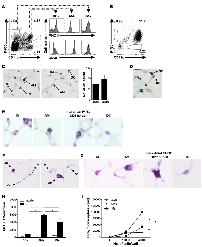
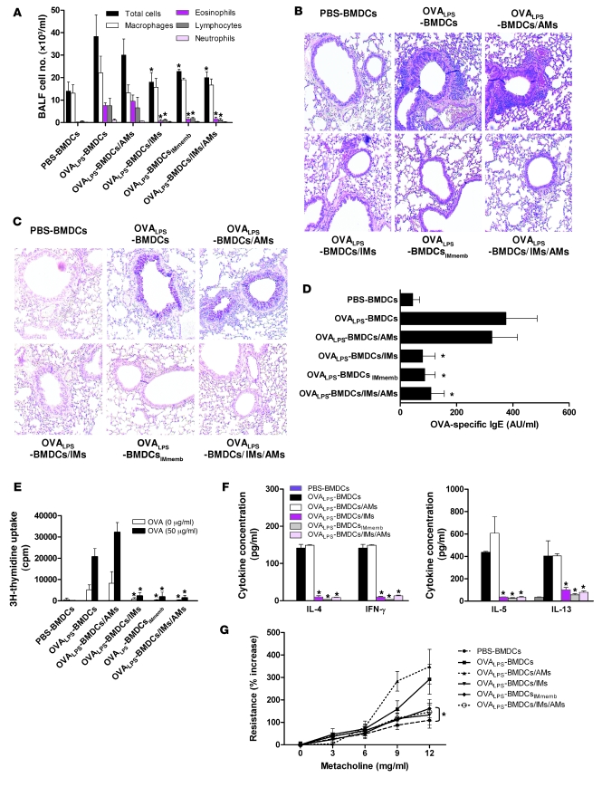
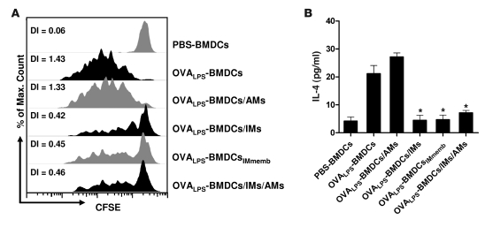
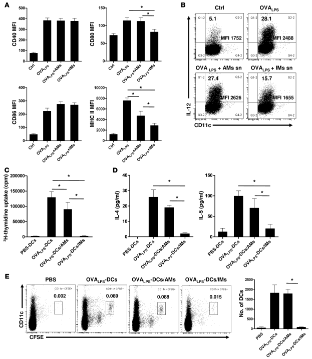
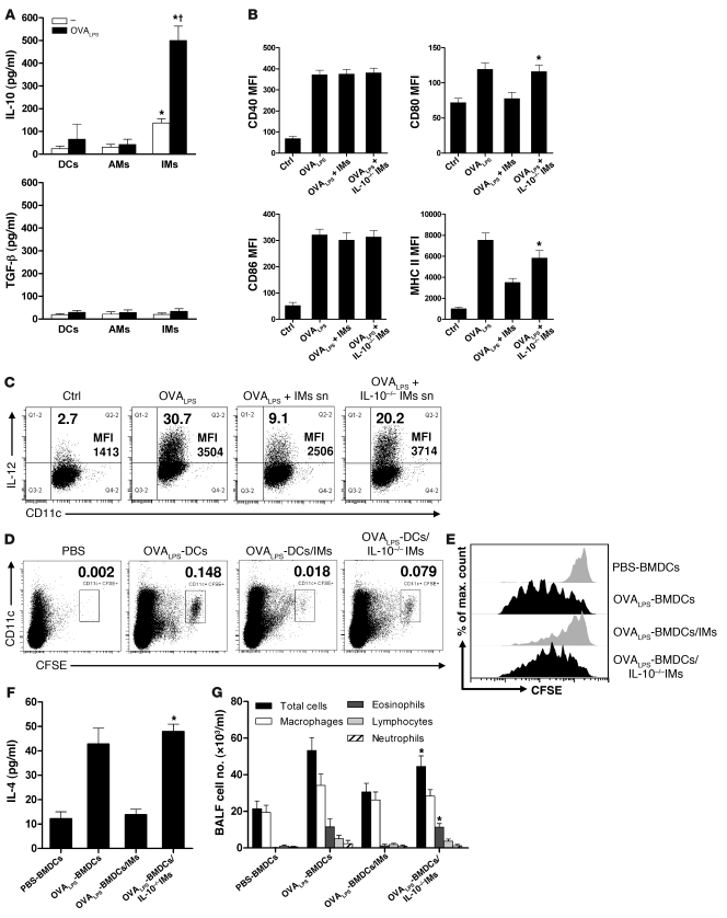
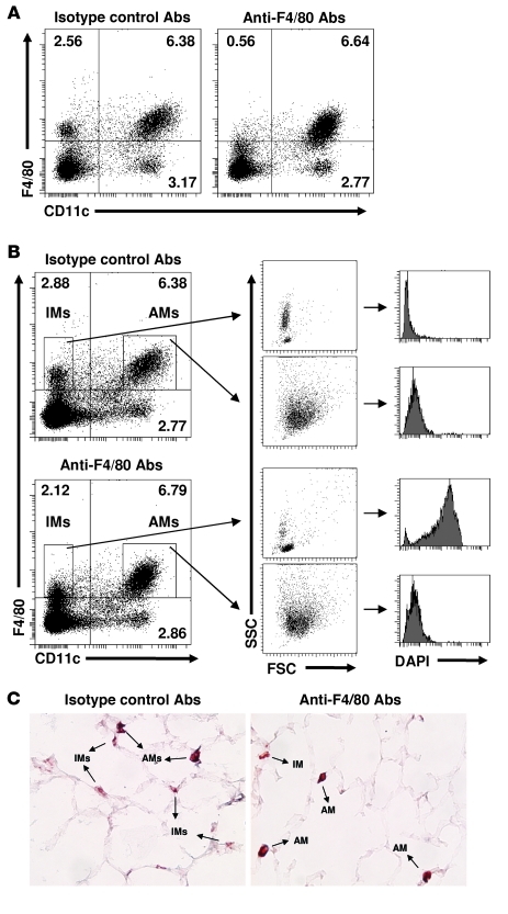
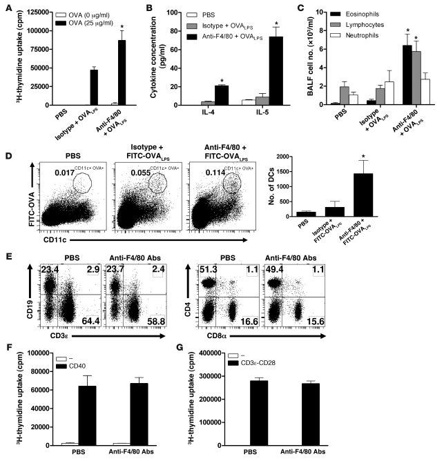
The respiratory tract is continuously exposed to both innocuous airborne antigens and immunostimulatory molecules of microbial origin, such as LPS. At low concentrations, airborne LPS can induce a lung DC-driven Th2 cell response to harmless inhaled antigens, thereby promoting allergic asthma. However, only a small fraction of people exposed to environmental LPS develop allergic asthma. What prevents most people from mounting a lung DC-driven Th2 response upon exposure to LPS is not understood. Here we have shown that lung interstitial macrophages (IMs), a cell population with no previously described in vivo function, prevent induction of a Th2 response in mice challenged with LPS and an experimental harmless airborne antigen. IMs, but not alveolar macrophages, were found to produce high levels of IL-10 and to inhibit LPS-induced maturation and migration of DCs loaded with the experimental harmless airborne antigen in an IL-10-dependent manner. We further demonstrated that specific in vivo elimination of IMs led to overt asthmatic reactions to innocuous airborne antigens inhaled with low doses of LPS. This study has revealed a crucial role for IMs in maintaining immune homeostasis in the respiratory tract and provides an explanation for the paradox that although airborne LPS has the ability to promote the induction of Th2 responses by lung DCs, it does not provoke airway allergy under normal conditions.


Figures

Figure 1
Figure 1. Phenotypic and functional comparison of IMs, AMs, and lung DCs.
(A) The percentage of IMs (F4/80+CD11c), AMs (F4/80+CD11c+), and DCs (F4/80CD11c+) in whole lung from BALB/c mice was determined by flow cytometry. IMs, AMs, and DCs were also stained for MHC II and CD68. (B) The percentage of F4/80+CD11c, F4/80+CD11c+, and F4/80CD11c+ cells in BALF from BALB/c mice. (CE) Lung cryosections were double stained for CD11c (blue) and F4/80 (red). IMs (F4/80+CD11c) are stained red, DCs (F4/80CD11c+) are stained blue, and AMs (F4/80+CD11c+) are double stained. (C) Representative photographs showing some IMs, AMs, and DCs (original magnification, ×100). The average number of IMs and AMs per field was calculated. (D) Image of IMs in the vicinity of DCs (original magnification, ×100). (E) Photographs at higher magnification showing IMs, AMs, interstitial F4/80+CD11c+ macrophages, and DCs (original magnification, ×200). (F and G) Alternatively, F4/80 was stained pink rather than red. (F) Representative photographs showing some IMs (pink), DCs (blue), and AMs (purple) (original magnification, ×100). (G) Photographs at higher magnification (original magnification, ×200). (H) FACS-sorted APCs were cocultured with FITC-labeled dextran in the absence or presence of sodium azide. After 45 minutes, the uptake of fluorescent dextran was determined by flow cytometry. (I) FACS-sorted APCs (1 × 10⁴ or 4 × 10⁴ cells/well) were loaded with the DO11.10 OVA peptide and cocultured for 3 days with naive DO11.10 CD4+ T cells. DO11.10 T cell proliferation was assessed by [3H]thymidine uptake in a 16-hour pulse. *P < 0.05 (H and I).
Figure 2
Figure 2. IMs are able to suppress the induction of airway allergy by OVA-pulsed, LPS-stimulated DCs.
(AG) Naive BALB/c mice were injected i.t. with PBS-BMDCs, OVALPS-BMDCs, OVALPS-BMDCs/AMs, OVALPS-BMDCs/IMs, OVALPS-BMDCsIMmemb, or OVALPS-BMDCs/IMs/AMs. From day 10 to day 14, mice were exposed to OVA aerosols. Twenty-four hours after the last challenge, the severity of airway allergy was evaluated. (A) BALF was subjected to total and differential cell counts. (B and C) Lung sections were stained with either H&E (B) or PAS (C) (original magnification, ×100). (D) Levels of OVA-specific IgE were measured in serum samples by ELISA. OVA-specific IgE levels are expressed as AU. (E and F) MLN cells were restimulated in vitro for 3 days with 50 μg/ml OVA. (E) The proliferation was measured as [3H]thymidine incorporation during the last 16 hours. (F) Culture supernatants were assayed for IL-4, IFN-γ, IL-5, and IL-13 by ELISA. (G) AHR to various doses of methacholine was assessed by invasive measurement of dynamic resistance. *P < 0.05 versus OVALPS-BMDCs and OVALPS-BMDCs/AMs (A and DG).
Figure 3
Figure 3. IMs can inhibit DC-mediated priming of Th2 cells.
(A and B) Naive BALB/c mice were injected i.v. with 107 CFSE-labeled DO11.10 T cells (day –1). Twenty-four hours later (day 0), mice received an i.t. administration of PBS-BMDCs, OVALPS-BMDCs, OVALPS-BMDCs/AMs, OVALPS-BMDCs/IMs, OVALPS-BMDCsIMmemb, or OVALPS-BMDCs/IMs/AMs. On day 3, MLNs were collected. (A) Proliferation of CFSE-labeled OVA-specific T cells was measured by flow cytometry. DI, division index; Max, maximum. (B) MLN cells were restimulated in vitro for 3 days with 25 μg/ml OVA, and supernatants were assayed for IL-4 by ELISA. *P < 0.05 versus OVALPS-BMDCs and OVALPS-BMDCs/AMs.
Figure 4
Figure 4. IMs have the capacity to attenuate LPS-induced maturation and migration of antigen-loaded DCs.
(A) Lung DCs (106 cells) from BALB/c mice were stimulated for 16 hours with OVALPS in the presence or absence of AMs (2 × 10⁶ cells) or IMs (1 × 10⁶ cells). DCs were then assayed for expression of CD40, CD80, CD86, and MHC II by FACS. Freshly isolated lung DCs served as control (Ctrl). MFIs are shown. (B) Lung DCs were placed in fresh medium or in the supernatant (sn) of OVALPS-stimulated IMs or AMs. DCs were then treated with OVALPS for 2 hours, incubated with Brefeldin A for an additional 5 hours, and finally stained for IL-12p40. The percentage of IL-12–positive cells was measured by FACS. (C and D) Lung DCs (2 × 10⁴ cells) were stimulated for 16 hours with OVALPS in the presence or absence of AMs (4 × 10⁴ cells) or IMs (2 × 10⁴ cells). APCs were then cocultured for 72 hours with 2 × 10⁵ DO11.10 CD4+ T cells. Unpulsed DCs were used as controls. (C) DO11.10 T cell proliferation was measured. (D) IL-4 and IL-5 were measured in the supernatants (ELISA). (E) CFSE-labeled OVALPS-DCs, OVALPS-DCs/AMs, or OVALPS-DCs/IMs were injected in the trachea of naive mice. Control mice received PBS. Twenty-four hours later, MLNs were digested and stained for F4/80 and CD11c. The percentages of migrating DCs (CFSE+F4/80CD11c+) among total MLN cells were determined by FACS. The total numbers of migrating DCs were calculated. *P < 0.05 (A and CE).
Figure 5
Figure 5. IL-10 secretion by IMs is required for functional paralysis of LPS-stimulated DCs.
(A) IMs, AMs, and lung DCs were stimulated with OVALPS. Sixteen hours later, supernatants were assayed for IL-10 and TGF-β. *P < 0.05 versus AMs and DCs. P < 0.05 versus unstimulated IMs. (B) Lung DCs were stimulated for 16 hours with OVALPS in the presence or absence of WT or Il10–/– IMs. DCs were assayed for expression of CD40, CD80, CD86, and MHC II by FACS. MFIs are shown. Ctrl, freshly isolated DCs. *P < 0.05 versus Ctrl and OVALPS plus IMs. (C) Lung DCs were placed in fresh medium or in the supernatant of OVALPS-stimulated WT or Il10–/– IMs. DCs were treated and stained as in Figure 4B. The percentage and MFI of IL-12–positive cells were measured by FACS. (D) CFSE-labeled OVALPS-DCs, OVALPS-DCs/IMs, and OVALPS-DCs/Il10–/– IMs were injected i.t. to recipients. Control mice received PBS. Twenty-four hours later, the percentages of migrating DCs (CFSE+F4/80CD11c+) among total MLN cells were determined by FACS. (E and F) Mice were injected with CFSE-labeled OT-II T cells. Twenty-four hours later, mice received PBS-BMDCs, OVALPS-BMDCs, OVALPS-BMDCs/IMs, or OVALPS-BMDCs/Il10–/– IMs. Three days later, proliferation of CFSE-labeled OVA-specific T cells in MLNs was measured by FACS (E). Alternatively, MLN cells were restimulated with OVA, and supernatants were assayed for IL-4 (F). (G) Mice received PBS-BMDCs, OVALPS-BMDCs, OVALPS-BMDCs/IMs, or OVALPS-BMDCs/IL-10–/– IMs. From days 10 to 14, mice were exposed to OVA aerosols. On day 15, BALF cell numbers were determined. *P < 0.05 versus PBS-BMDCs and OVALPS-BMDCs/IMs (F and G).
Figure 6
Figure 6. Specific depletion of IMs by i.p. injection of anti-F4/80 antibodies.
(AC) Naive BALB/c mice were injected i.p. on 3 consecutive days with 250 μg of depleting anti-F4/80 or control isotype antibodies. (A and B) On day 4, lungs were digested and stained for F4/80 and CD11c. (A) Percentages of lung DCs (F4/80CD11c+; lower-right quadrant), AMs (F4/80+CD11c+; upper-right), and IMs (F4/80+CD11c; upper-left) among living cells. (B) Percentages of lung DCs, AMs, and IMs among dead and living cells (all events were considered; left panels). FACS analysis of the forward scatter (FSC) and side scatter (SSC) is provided to show the specific effects of depleting anti-F4/80 antibodies on IM morphology (middle panels). Cells were incubated with DAPI in order to stain dead cells (right panels). Only IMs were dead (DAPI-positive) following anti-F4/80 treatment. (C) Lung cryosections from isotype antibody– and anti-F4/80 antibody–treated mice were stained for the pan-macrophage marker CD68 (original magnification, ×100).
Figure 7
Figure 7. IMs prevent LPS-triggered Th2 responses to innocuous inhaled antigens.
(AG) Naive BALB/c mice were injected i.p. daily from day 1 to 3 with depleting anti-F4/80 or control isotype antibodies. (AC) On day 2, mice received an i.t. injection of OVALPS. (A and B) On day 6, MLN cells were restimulated for 3 days with 25 μg/ml OVA. The proliferation was measured (A), and culture supernatants were assayed for IL-4 and IL-5 by ELISA (B). (C) From day 11 to 14, mice were challenged intranasally with 25 μg OVA (grade V; Sigma-Aldrich) in 50 μl PBS. On day 15, BALF was subjected to differential cell counts. (D) On day 2, IM-depleted and control mice were injected i.t. with 100 μg FITC-OVA (rather than unlabeled OVA) and 10 ng LPS. On day 3, MLNs were analyzed by flow cytometry for the presence of OVA-loaded DCs (FITC+F4/80CD11c+). Percentages (left) and total numbers (right) of migrating DCs are shown. (E) On day 4, the percentages of T (CD3ε+, CD4+, or CD8α+ cells) and B (CD19+ cells) cells in MLNs were measured by flow cytometry. (F and G) On day 4, B and T cells were isolated from MLNs and stimulated ex vivo with agonist anti-CD40 antibodies or anti-CD3 and anti-CD28 antibodies, respectively. Control cells were left unstimulated. B cell (F) and T cell (G) proliferation was measured as [3H]thymidine incorporation during the last 16 hours of a 2-day culture. *P < 0.05 versus results obtained with isotype control antibodies (AD).

Similar articles

Cited by

References

    1. Akbari O., DeKruyff R., Umetsu D. Pulmonary dendritic cells producing IL-10 mediate tolerance induced by respiratory exposure to antigen. Nat. Immunol. 2001;2:725–731. doi: 10.1038/90667. - DOI - PubMed
    1. Akbari O., et al. Antigen-specific regulatory T cells develop via the ICOS-ICOS-ligand pathway and inhibit allergen-induced airway hyperreactivity. Nat. Med. 2002;8:1024–1032. doi: 10.1038/nm745. - DOI - PubMed
    1. de Heer H., et al. Essential role of lung plasmacytoid dendritic cells in preventing asthmatic reactions to harmless inhaled antigen. J. Exp. Med. 2004;200:89–98. doi: 10.1084/jem.20040035. - DOI - PMC - PubMed
    1. Michel O., et al. Severity of asthma is related to endotoxin in house dust. Am. J. Respir. Crit. Care Med. 1996;154:1641–1646. - PubMed
    1. Milton D., Johnson D., Park J. Environmental endotoxin measurement: interference and sources of variation in the Limulus assay of house dust. Am. Ind. Hyg. Assoc. J. 1997;58:861–867. - PubMed

Publication types

MeSH terms