. 2009 Nov 12;4(11):e7787.
doi: 10.1371/journal.pone.0007787.

Diminished macrophage apoptosis and reactive oxygen species generation after phorbol ester stimulation in Crohn's disease

Affiliations

Diminished macrophage apoptosis and reactive oxygen species generation after phorbol ester stimulation in Crohn's disease

Christine D Palmer et al. PLoS One. .

Abstract

Background: Crohn's Disease (CD) is a chronic relapsing disorder characterized by granulomatous inflammation of the gastrointestinal tract. Although its pathogenesis is complex, we have recently shown that CD patients have a systemic defect in macrophage function, which results in the defective clearance of bacteria from inflammatory sites.

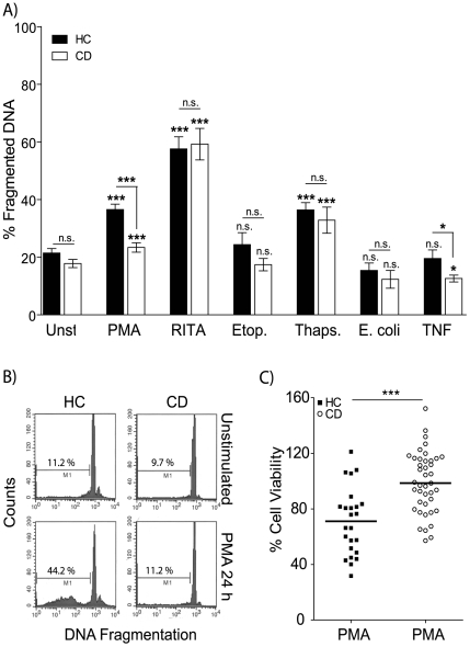
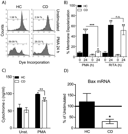
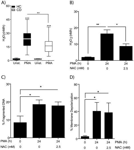
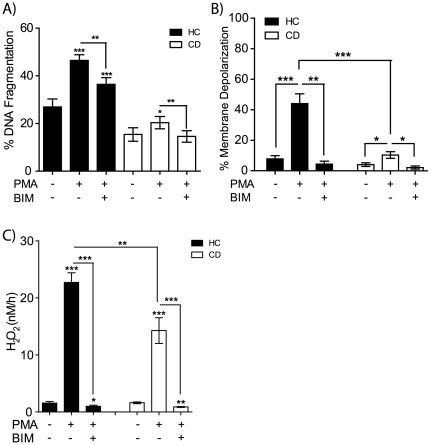
Methodology/principal findings: Here we have identified a number of additional macrophage defects in CD following diacylglycerol (DAG) homolog phorbol-12-myristate-13-acetate (PMA) activation. We provide evidence for decreased DNA fragmentation, reduced mitochondrial membrane depolarization, impaired reactive oxygen species production, diminished cytochrome c release and increased IL-6 production compared to healthy subjects after PMA exposure. The observed macrophage defects in CD were stimulus-specific, as normal responses were observed following p53 activation and endoplasmic reticulum stress.

Conclusion: These findings add to a growing body of evidence highlighting disordered macrophage function in CD and, given their pivotal role in orchestrating inflammatory responses, defective apoptosis could potentially contribute to the pathogenesis of CD.


Conflict of interest statement

Competing Interests: The authors have declared that no competing interests exist.

Figures

Figure 1
Figure 1. Defective apoptosis in macrophages from CD subjects.
A Macrophages from HC and CD patients were untreated or stimulated with 1 µg/ml PMA (HC n = 39, CD n = 44), 1 µM RITA (HC n = 10, CD n = 7), 25 µM Etoposide (HC n = 7, CD n = 7), 1 µM Thapsigargin (HC n = 9, CD n = 8) for 24 h, E. coli (HC n = 10, CD n = 12) or 25 ng/ml recombinant TNF. DNA fragmentation (events below the G1-0 peak) was assessed by flow cytometry. Mean percentage ± SEM of apoptosis for HC (black bars) and CD (open bars) are shown (o significance between stimulated and untreated, * between HC and CD). B Representative histograms for HC unstimulated (upper left panel), HC plus PMA 24 h (lower left), CD unstimulated (upper right) and CD plus PMA 24 h (lower right). C Viability for CD and HC macrophages following stimulation with 1 µg/ml PMA for 24 h was assessed by MTT assay. Data are presented as percent of untreated cells for HC (black squares, n = 24) and CD (open circles, n = 41) with group mean. Statistical analysis: Paired or unpaired t-test. Symbols: p<0.05 (* or o), p<0.001 (*** or ooo).
Figure 2
Figure 2. Mitochondrial membrane depolarization, cytochrome c and BAX expression are abnormal in CD macrophages after PMA activation.
Macrophages were stimulated with 1 µg/ml PMA for 24 h and the effects on mitochondrial membrane potential, cytochrome c and BAX expression determined. A Representative histograms of macrophage population for HC unstimulated (upper left panel), HC PMA 24 h (lower left), CD unstimulated (upper right) and CD PMA 24 h (lower right) are shown. Gated populations show cells with an intact mitochondrial membranes (M1) and cells which have lost mitochondrial membrane integrity (M2). B Proportion of macrophages with mitochondrial membrane depolarization are shown as mean percentage ± SEM after either PMA (HC n = 9, CD n = 10) or RITA (HC n = 5, CD n = 5) stimulation. C Intracellular cytochrome C levels in macrophages stimulated with 1 µg/ml PMA for 24 h were measured by ELISA. Cytochrome C production (ng/ml) at 24 h is shown for HC (n = 4, black bars) and CD (n = 8, open bars). D Macrophages were stimulated with 1 µg/ml PMA for 4 h followed by total RNA isolation. Bax mRNA levels were determined by qPCR and expressed as the change in expression compared to unstimulated cells. Statistical analysis: Paired or unpaired t-test. Symbols: p<0.05 (*), p<0.01 (**), p<0.001 (***).
Figure 3
Figure 3. Decreased PMA-induced reactive oxygen species production in CD macrophages.
NADPH oxidase activity was assessed by measuring the generation of H2O2 by macrophages from HC and CD subjects after stimulation with 1 µg/ml PMA. A Production of H2O2 (nM/h) was elevated after PMA stimulation in both HC (n = 29) and CD (n = 47) groups. Macrophages from CD patients demonstrated reduced H2O2 release than HC. B H2O2 levels were determined after HC macrophages (n = 4) pre-incubated with 2.5 mM N-Acetyl-L-cysteine (NAC) for 1 h followed by PMA stimulation. The presence of NAC resulted in reduced H2O2 levels. C DNA fragmentation and D mitochondrial membrane potential were unaltered by the presence of NAC. Statistical analysis: Paired or unpaired t-test. Symbols: p<0.05 (*), p<0.01 (**), p<0.001 (***), HC (black bars), CD (white bars).
Figure 4
Figure 4. Dysregulated PMA-induced IL-6 production in CD macrophages.
A Cytokine secretion from HC (n = 7) and CD (n = 10) macrophages following stimulation with 1 µg/ml PMA for six hours was assessed. IL-6 production was significantly elevated in macrophages from CD patients compared to HC subjects. B Measuring the effect of elevated IL-6 (1 ng/ml) on DNA fragmentation in the presence or absence of PMA stimulation. DNA fragmentation was unaltered by the inclusion of IL-6 in both HC (n = 7) and CD (n = 7) macrophages. Statistical analysis: Paired or unpaired t-test. Symbols: p<0.05 (*), p<0.01 (**), p<0.001 (***).
Figure 5
Figure 5. PMA induced apoptosis, NADPH oxidase activity and mitochondrial membrane depolarization are all inhibited by Bisindolylmaleimide I.
Macrophages from HC (n = 5–10) and CD (n = 5–10) were left untreated, or pre-incubated with 1 µM Bisindolylmaleimide I (BIM) for 1 h followed by PMA stimulation. BIM significantly reduced A DNA fragmentation, B mitochondrial membrane depolarization and C H2O2 release in both HC and CD. Paired or unpaired t-test. Symbols: p<0.05 (*), p<0.01 (**), p<0.001 (***), HC (black bars), CD (white bars).

References

    1. Podolsky DK. Inflammatory bowel disease. N Engl J Med. 2002;347:417–429. - PubMed
    1. Shanahan F. Crohn's disease. Lancet. 2002;359:62–69. - PubMed
    1. Korzenik JR. Past and current theories of etiology of IBD: toothpaste, worms, and refrigerators. J Clin Gastroenterol. 2005;39:S59–S65. - PubMed
    1. Marks DJ, Harbord MW, MacAllister R, Rahman FZ, Young J, et al. Defective acute inflammation in Crohn's disease: a clinical investigation. Lancet. 2006;367:668–678. - PubMed
    1. Smith AM, Rahman FZ, Hayee B, Graham SJ, Marks DJ, et al. Disordered macrophage cytokine secretion underlies impaired acute inflammation and bacterial clearance in Crohn's disease. J Exp Med. 2009;206:1883–1897. - PMC - PubMed

Publication types