. 2009 Sep;2(5):444-7.
doi: 10.4161/cib.2.5.8931.

Endosomal recycling regulation during cytokinesis

Affiliations

Endosomal recycling regulation during cytokinesis

Erkang Ai et al. Commun Integr Biol. 2009 Sep.

Abstract

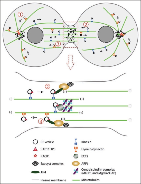
Successful cytokinesis is critical for cell proliferation and development. In animal cells, cytokinesis relies on temporally and spatially regulated membrane addition to the cleavage site. An important source for the new membrane is recycling endosomes. Yet how these endocytic vesicles are transported and regulated remains unclear. Several potential factors have been recently identified that regulate the trafficking of recycling endosomes during cytokinesis. Dynein and dynactin are required for the retrograde transport of recycling endosomes, while Kinesin-1 is responsible for endosome delivery to the furrow and midbody. Other regulators of recycling endosome trafficking have been identified, including RACK1, JIP3/4 and ECT2, which target recycling endosomes during the cell cycle. Here, we provide insights into the mechanisms controlling endosomal trafficking during cytokinesis.

Keywords: ARF6; JIP4; RAB11; RAB11FIP3; RACK1; cytokinesis; dynactin; dynein; kinesin; membrane trafficking; recycling endosomes.


Figures

Figure 1
Figure 1
Model of recycling endosome (RE) trafficking during cytokinesis. (1) REs are clustered at the MTOC. The endosomes are transported along the microtubules via dynein/dynactin motors. RACK1 at the centrosomes anchors dynactin and targets REs to the centrosomes. (2) RE vesicles are directed to the cleavage site via kinesin motors. They are trafficked along both spindle microtubules and midzone microtubules. At the cleavage site these vesicles interact with ARF6 and the exocyst complex and insert membrane into the plasma membrane and the midbody. (3) RE vesicles may also be transported away from the cleavage site by dynein/dynactin. ARF6 and JIP3/4 likely serve as the switch controlling whether RE vesicles associate with kinesin or dynein.

Similar articles

Cited by

References

    1. Bluemink JG, de Laat SW. New membrane formation during cytokinesis in normal and cytochalasin B-treated eggs of Xenopus laevis I. Electron microscope observations. J Cell Biol. 1973;59:89–108. - PMC - PubMed
    1. Prekeris R, Gould GW. Breaking up is hard to do—membrane traffic in cytokinesis. J Cell Sci. 2008;121:1569–1576. - PMC - PubMed
    1. Strickland LI, Burgess DR. Pathways for membrane trafficking during cytokinesis. Trends Cell Biol. 2004;14:115–118. - PubMed
    1. Staehelin LA, Hepler PK. Cytokinesis in higher plants. Cell. 1996;84:821–824. - PubMed
    1. Echard A, Hickson GR, Foley E, O'Farrell PH. Terminal cytokinesis events uncovered after an RNAi screen. Curr Biol. 2004;14:1685–1693. - PMC - PubMed

LinkOut - more resources