

Differentiation of neural precursors and dopaminergic neurons from human embryonic stem cells
- PMID: 19907987
- PMCID: PMC2948208
- DOI: 10.1007/978-1-60761-369-5_19
Differentiation of neural precursors and dopaminergic neurons from human embryonic stem cells
Abstract
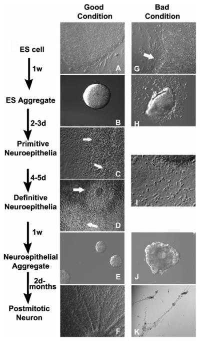
Directed differentiation of human embryonic stem cells (hESCs) to a functional cell type, including neurons, is the foundation for application of hESCs. We describe here a reproducible, chemically defined protocol that allows directed differentiation of hESCs to nearly pure neuroectodermal cells and neurons. First, hESC colonies are detached from mouse fibroblast feeder layers and form aggregates to initiate the differentiation procedure. Second, after 4 days of suspension culture, the ESC growth medium is replaced with neural induction medium to guide neuroectodermal specification. Third, the differentiating hESC aggregates are attached onto the culture surface at day 6-7, where columnar neural epithelial cells appear and organize into rosettes. Fourth, the neural rosettes are enriched by detaching rosettes and leaving the peripheral flat cells attached and expanded as neuroepithelial aggregates in the same medium. Finally, the neuroepithelial aggregates are dissociated and differentiated to nearly pure neurons. This stepwise differentiation protocol results in the generation of primitive neuroepithelia at day 8-10, neural progenitors at the second and third week, and postmitotic neurons at the fourth week, which mirrors the early phase of neural development in a human embryo. Identification of the primitive neuroepithelial cells permits efficient patterning of region-specific progenitors and neuronal subtypes such as midbrain dopaminergic neurons.
Figures
References
-
- Zhang SC. Embryonic stem cells for neural replacement therapy: Prospects and Challenges. J. Hematother. Stem Cell Res. 2003;12:625–634. - PubMed
-
- Du ZW, Zhang SC. Neural differentiation from embryonic stem cells: which way? Stem Cell Development. 2004;13:372–381. - PubMed
-
- Zhang SC, Wernig M, Duncan ID, Brüstle O, Thomson JA. In vitro differentiation of transplantable neural precursors from human embryonic stem cells. Nat. Biotechnol. 2001;19:1129–1133. - PubMed
-
- Hemmati-Brivanlou A, Melton D. Vertebrate embryonic cells will become nerve cells unless told otherwise. Cell. 1997;88:13–17. - PubMed
Publication types
MeSH terms
Substances
Grants and funding
LinkOut - more resources
Full Text Sources
Other Literature Sources
