

doi: 10.1007/978-1-60761-380-0_29.
Limiting dilution analysis of murine epidermal stem cells using an in vivo regeneration assay
Affiliations
- PMID: 19908020
- PMCID: PMC2783164
- DOI: 10.1007/978-1-60761-380-0_29
Item in Clipboard
Limiting dilution analysis of murine epidermal stem cells using an in vivo regeneration assay
Methods Mol Biol.
2010.
Abstract
Epidermal stem cells are of major importance for tissue homeostasis, wound repair, tumor initiation, and gene therapy. Here we describe an in vivo regeneration assay to test for the ability of keratinocyte progenitors to maintain an epidermis over the long-term in vivo. Limiting dilution analysis of epidermal repopulating units in this in vivo regeneration assay at sequential time points allows the frequency of short-term (transit amplifying cell) and long-term (stem cell) repopulating cells to be quantified.
Figures
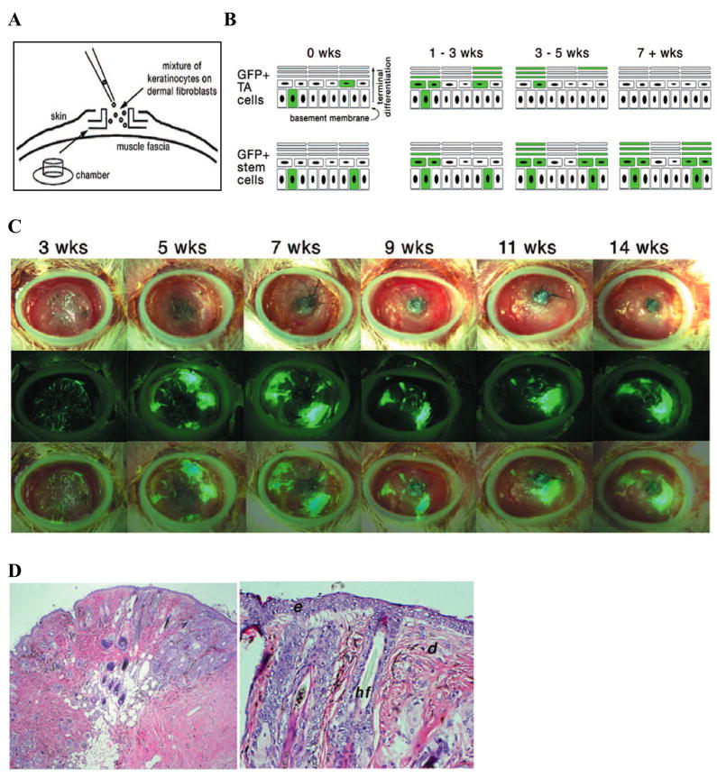
Limiting dilution analysis for the determination of short term (TA) and long term (stem) repopulating cell frequency. (A) Diagram of the 6 mm diameter silicone chamber implanted onto the dorsal fascia of a NODSCID mouse into which the keratinocyte and fibroblast populations are seeded. (B) Schematic of epidermal layers where GFP positive units derived from short term repopulating cells (TA cells) disappear after 3 to 5 weeks (top row) or GFP positive cells derived from long term repopulating cells (stem cells) persist for 9 to 30 weeks. (C) Maintenance of GFP positive repopulating units up to 14 weeks. Top panels show bright-field images of regenerated skin, middle panels show epifluorescence images taken at the same time, and bottom panels show an overlay of the GFP positive repopulating units on the regenerated skin. (D) Hematoxylin and eosin staining of regenerated skin in cross section (10x, 20x). d: dermis, e: epidermis, hf: hair follicle.
Similar articles
-
CD133 is a marker for long-term repopulating murine epidermal stem cells.J Invest Dermatol. 2012 Nov;132(11):2522-33. doi: 10.1038/jid.2012.196. Epub 2012 Jul 5. J Invest Dermatol. 2012. PMID: 22763787 Free PMC article.
-
The CD44+ ALDH+ population of human keratinocytes is enriched for epidermal stem cells with long-term repopulating ability.Stem Cells. 2013 Apr;31(4):786-99. doi: 10.1002/stem.1329. Stem Cells. 2013. PMID: 23335266 Free PMC article.
-
Competitive Repopulation Assay of Long-Term Epidermal Stem Cell Regeneration Potential.Methods Mol Biol. 2020;2109:45-53. doi: 10.1007/7651_2019_234. Methods Mol Biol. 2020. PMID: 31087286
-
Interfollicular epidermal stem cells: identification, challenges, potential.J Invest Dermatol. 2006 Jul;126(7):1450-8. doi: 10.1038/sj.jid.5700184. J Invest Dermatol. 2006. PMID: 16543901 Review.
-
Interfollicular epidermal homeostasis: dicing with differentiation.Exp Dermatol. 2012 Apr;21(4):249-53. doi: 10.1111/j.1600-0625.2012.01447.x. Exp Dermatol. 2012. PMID: 22417300 Review.
Cited by
-
ABCG2-dependent dye exclusion activity and clonal potential in epithelial cells continuously growing for 1 month from limbal explants.Invest Ophthalmol Vis Sci. 2011 Jun 17;52(7):4330-7. doi: 10.1167/iovs.10-5897. Invest Ophthalmol Vis Sci. 2011. PMID: 21421882 Free PMC article.
-
Establishment of a murine epidermal cell line suitable for in vitro and in vivo skin modelling.BMC Dermatol. 2011 Apr 21;11:9. doi: 10.1186/1471-5945-11-9. BMC Dermatol. 2011. PMID: 21510892 Free PMC article.
References
-
- Hodgson GS, Bradley TR. Properties of haematopoietic stem cells surviving 5-fluorouracil treatment: evidence for a pre-CFU-S cell? Nature. 1979;281(5730):381–2. - PubMed
-
- Lerner C, Harrison DE. 5-Fluorouracil spares hemopoietic stem cells responsible for long-term repopulation. Exp Hematol. 1990;18(2):114–8. - PubMed
MeSH terms
Grants and funding
LinkOut - more resources
Full Text Sources
Medical
