Case Reports
. 2010 Apr;33(2):355-61.
doi: 10.1007/s00270-009-9727-z. Epub 2009 Nov 12.

Prostatic artery embolization as a primary treatment for benign prostatic hyperplasia: preliminary results in two patients

Affiliations
Case Reports

Prostatic artery embolization as a primary treatment for benign prostatic hyperplasia: preliminary results in two patients

Francisco Cesar Carnevale et al. Cardiovasc Intervent Radiol. 2010 Apr.

Abstract

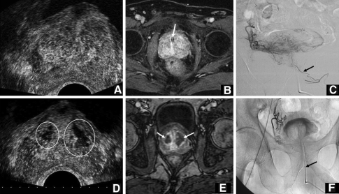
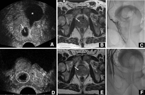
Symptomatic benign prostatic hyperplasia (BPH) typically occurs in the sixth and seventh decades, and the most frequent obstructive urinary symptoms are hesitancy, decreased urinary stream, sensation of incomplete emptying, nocturia, frequency, and urgency. Various medications, specifically 5-alpha-reductase inhibitors and selective alpha-blockers, can decrease the severity of the symptoms secondary to BPH, but prostatectomy is still considered to be the traditional method of management. We report the preliminary results for two patients with acute urinary retention due to BPH, successfully treated by prostate artery embolization (PAE). The patients were investigated using the International Prostate Symptom Score, by digital rectal examination, urodynamic testing, prostate biopsy, transrectal ultrasound (US), and magnetic resonance imaging (MRI). Uroflowmetry and postvoid residual urine volume complemented the investigation at 30, 90, and 180 days after PAE. The procedure was performed under local anesthesia; embolization of the prostate arteries was performed with a microcatheter and 300- to 500-microm microspheres using complete stasis as the end point. One patient was subjected to bilateral PAE and the other to unilateral PAE; they urinated spontaneously after removal of the urethral catheter, 15 and 10 days after the procedure, respectively. At 6-month follow-up, US and MRI revealed a prostate reduction of 39.7% and 47.8%, respectively, for the bilateral PAE and 25.5 and 27.8%, respectively, for the patient submitted to unilateral PAE. The early results, at 6-month follow-up, for the two patients with BPH show a promising potential alternative for treatment with PAE.


Figures

Fig. 1
Fig. 1
Patient 1. A Transversal view of the prostate gland. Baseline pre-embolization contrast-enhanced ultrasound. Homogeneous enhancement of the internal gland. B Pre-embolization MRI. Axial postcontrast T1-weighted MRI depicting the enlarged prostate due to central gland nodules. Note the presence of the urethral catheter (white arrow). C Angiogram pre-embolization showing the right inferior vesical artery and the right prostate branches. The branches to the prostate communicate with the corresponding vessels of the opposite side (black arrow). D Six-month follow-up after PAE contrast-enhanced ultrasound. Normal enhancement in the hyperechoic areas of internal gland and hypoechoic avascular areas (white circles). E Six-month postembolization MRI. Axial postcontrast T1-weighted MRI depicting bilateral avascular areas in the central gland (white arrows) and a reduction in prostate size. F Angiogram after right inferior vesical artery and right prostate branch embolization. Transurethral catheter in place (black arrow)
Fig. 2
Fig. 2
Patient 2. A Baseline pre-embolization contrast-enhanced ultrasound. Tranverse view of the prostate gland. Asymmetric enlargement of the internal gland. Homogeneous enhancement of the internal gland characterized by hyperechoic areas. Note the small cyst in the right internal gland (white arrow) and the Foley catheter (*). B Pre-embolization MRI. Axial T2-weighted MRI depicting an enlarged prostate due to nodules in the central gland, with a more cystic nodule on the right side (white arrow). Note the presence of the urethral catheter (black arrow). C Angiogram pre-embolization showing the right inferior vesical artery and the right prostate branches delimiting the right side of the gland. D Six-month follow-up after PAE. Contrast-enhanced ultrasound shows normal enhancement in the hyperechoic areas of the internal gland and hypoechoic avascular area (white circle). E Six-month postembolization MRI. Axial T2-weighted MRI depicting a reduction in prostate size and the persistence of a conspicuous cystic nodule on the right side (white arrow). F Angiogram control after right inferior vesical artery and right prostate branch embolization

Similar articles

Cited by

References

    1. Emberton M, Andriole GL, la Rosette J, et al. Benign prostatic hyperplasia. A progressive disease of aging men. Urology. 2003;61:267–273. doi: 10.1016/S0090-4295(02)02371-3. - DOI - PubMed
    1. Ziada A, Rosenblum M, Crawford ED. Benign prostatic hyperplasia: an overview. Urology. 1999;53(3; Suppl 3a):1–6. doi: 10.1016/S0090-4295(98)00532-9. - DOI - PubMed
    1. Guess HA, Arrighi HM, Metter EJ, et al. Cumulative prevalence of prostatism matches the autopsy prevalence of benign prostatic hyperplasia. Prostate. 1990;17(3):241–246. doi: 10.1002/pros.2990170308. - DOI - PubMed
    1. AUA Practice Guidelines Committee AUA guideline on management of benign prostatic hyperplasia. I. Diagnosis and treatment recommendations. J Urol. 2003;170(2; pt 1):530–547. - PubMed
    1. Roehrborn CG, Preminger G, Newhall P, et al. Microwave thermotherapy for benign prostatic hyperplasia with the Dornier urowave: results of a randomized, double-blind, multicenter, sham-controlled trial. Urology. 1998;51(1):19–28. doi: 10.1016/S0090-4295(97)00571-2. - DOI - PubMed

Publication types

MeSH terms