Review
. 2009 Dec;19(4):436-50.
doi: 10.1007/s11065-009-9118-x. Epub 2009 Nov 12.

Beta-amyloid deposition and the aging brain

Affiliations
Review

Beta-amyloid deposition and the aging brain

Karen M Rodrigue et al. Neuropsychol Rev. 2009 Dec.

Abstract

A central issue in cognitive neuroscience of aging research is pinpointing precise neural mechanisms that determine cognitive outcome in late adulthood as well as identifying early markers of less successful cognitive aging. One promising biomarker is beta amyloid (Abeta) deposition. Several new radiotracers have been developed that bind to fibrillar Abeta providing sensitive estimates of amyloid deposition in various brain regions. Abeta imaging has been primarily used to study patients with Alzheimer's Disease (AD) and individuals with Mild Cognitive Impairment (MCI); however, there is now building data on Abeta deposition in healthy controls that suggest at least 20% and perhaps as much as a third of healthy older adults show significant deposition. Considerable evidence suggests amyloid deposition precedes declines in cognition and may be the initiator in a cascade of events that indirectly leads to age-related cognitive decline. We review studies of Abeta deposition imaging in AD, MCI, and normal adults, its cognitive consequences, and the role of genetic risk and cognitive reserve.


Figures

Fig. 1
Fig. 1
Illustration of beta-amyloid plaques in fixed tissue at autopsy (from Josephs et al. 2008). a shows an example of sparse Aβ burden; b illustrates a brain with moderate Aβ; and c is an example of frequent Aβ deposition in the brain
Fig. 2
Fig. 2
18F AV-45 uptake in an AD subject (left panel top) and a Healthy Control (left panel bottom). The right panel displays normalized signal uptake value (SUVr) in patients with AD and healthy controls in the precuneus and neocortex
Fig. 3
Fig. 3
An illustration of the timecourse of the rate of the decay of a radiotracer. As each half-life is passed the radioactive material is reduced by 50% and follows an exponential function
Fig. 4
Fig. 4
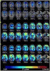
Illustration of typical Aβ deposition across varying groups and stages of pathology. Examples are from left to right, images of a PIB negative healthy older adult, a PIB positive healthy older adult, a PIB negative MCI subject, a PIB positive MCI subject, and an AD patient. Blues and purples represent little Aβ deposition and yellow and red the highest deposition (from Pike et al. 2007)
Fig. 5
Fig. 5
Illustration of association between APOE genotype and Aβ deposition in the brain in cognitively normal older adults (from Reiman et al. 2009). a displays a small to moderate increase in PIB binding in the individuals heterozygous for ε4 compared with non-carriers; b illustrates the greater increased PIB binding in individuals homozygous for ε4 compared with non-carriers; c depicts the association of greater PIB binding and APOE- ε4 dose for the whole sample
Fig. 6
Fig. 6
Illustration of normal variation in magnitude and extent of Aβ deposition in healthy controls (from Jack et al. 2008). a is the scan from the subject with the highest PIB retention in the study (despite being a normal control and cognitively normal) and b is the scan from a healthy control subject with low PIB retention, who was cognitively normal, but scored lower than the subject depicted in panel (a)
Fig. 7
Fig. 7
Proposed model relating imaging, pathology and clinical presentation over an individual’s adult lifetime (from Jack et al. 2009). The lifetime course of progression from presymptomatic, prodromal (MCI), and dementia (AD) phases is plotted. Neurodegeneration, detected by MRI, is indicated by a dashed line. Cognitive function is indicated by a dot-dash line. Amyloid deposition is indicated by a solid line late in life (i.e. that portion of the disease for which data currently exist). The time course of amyloid deposition early in life is represented as two possible theoretical trajectories (dotted lines), reflecting uncertainty about the time course trajectory of early Aβ deposition
Fig. 8
Fig. 8
A conceptual model of the scaffolding theory of aging and cognition (STAC; adapted from Park and Reuter-Lorenz 2009)

References

    1. Aizenstein HJ, Nebes RD, Saxton JA, Price JC, Mathis CA, Tsopelas ND, et al. Frequent amyloid deposition without significant cognitive impairment among the elderly. Archives of Neurology. 2008;65:1509–1517. - PMC - PubMed
    1. Andreasen N, Minthon L, Vanmechelen E, Vanderstichele H, Davidsson P, Winblad B, et al. Cerebrospinal fluid tau and Abeta42 as predictors of development of Alzheimer’s disease in patients with mild cognitive impairment. Neuroscience Letters. 1999;273:5–8. - PubMed
    1. Andrews-Hanna JR, Snyder AZ, Vincent JL, Lustig C, Head D, Raichle ME, et al. Disruption of large-scale brain systems in advanced aging. Neuron. 2007;56:924–935. - PMC - PubMed
    1. Bennett DA, Schneider JA, Arvanitakis Z, Kelly JF, Aggarwal NT, Shah RC, et al. Neuropathology of older persons without cognitive impairment from two community-based studies. Neurology. 2006;66:1837–1844. - PubMed
    1. Bourgeat P, Villemagne VL, Fripp J, Pike KE, Raniga P, Acosta O, et al. Relation between amyloid burden, brain atrophy and memory in Alzheimer’s disease. Alzheimer’s Association 2009 International Conference on Alzheimer’s Disease (ICAD); 2009, July.2009.

Publication types

Substances