. 2010 May 1;88(6):1182-92.
doi: 10.1002/jnr.22288.

Promoting directional axon growth from neural progenitors grafted into the injured spinal cord

Affiliations

Promoting directional axon growth from neural progenitors grafted into the injured spinal cord

Joseph F Bonner et al. J Neurosci Res. .

Abstract

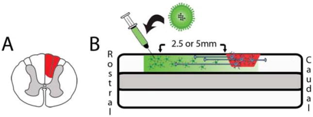
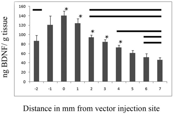
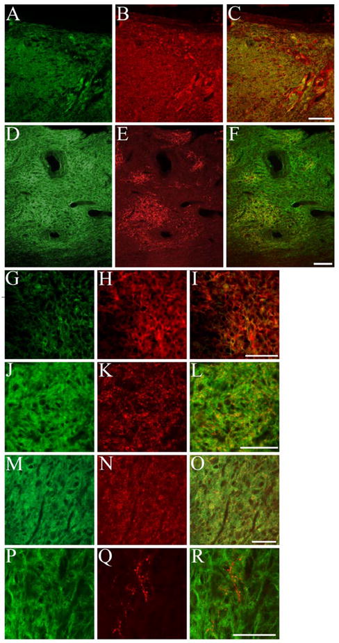
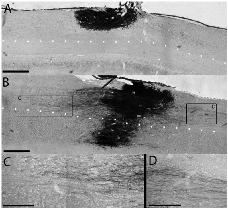
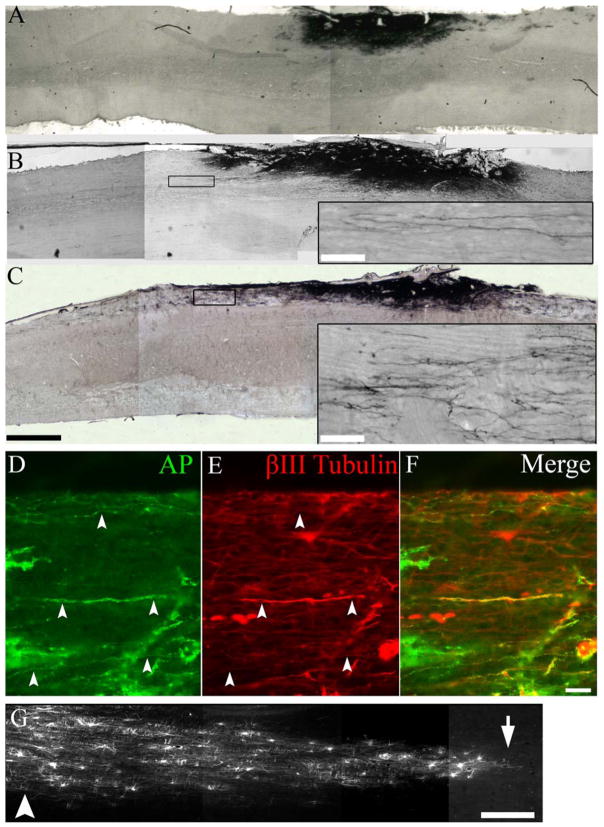
Spinal cord injury (SCI) is a devastating condition characterized by disruption of axonal connections, failure of axonal regeneration, and loss of motor and sensory function. The therapeutic promise of neural stem cells has been focused on cell replacement, but many obstacles remain in obtaining neuronal integration following transplantation into the injured CNS. This study investigated the neurotransmitter identity and axonal growth potential of neural progenitors following grafting into adult rats with a dorsal column lesion. We found that using a combination of neuronal and glial restricted progenitors (NRP and GRP) produced graft-derived glutamatergic and GABAergic neurons within the injury site, with minimal axonal extension. Administration of brain-derived neurotrophic factor (BDNF) with the graft promoted modest axonal growth from grafted cells. In contrast, injecting a lentiviral vector expressing BDNF rostral into the injured area generated a neurotrophin gradient and promoted directional growth of axons for up to 9 mm. Animals injected with BDNF lentivirus (at 2.5 and 5.0 mm) showed significantly more axons and significantly longer axons than control animals injected with GFP lentivirus. However, only the 5.0-mm-BDNF group showed a preference for extension in the rostral direction. We concluded that NRP/GRP grafts can be used to produce excitatory and inhibitory neurons, and neurotrophin gradients can guide axonal growth from graft-derived neurons toward putative targets. Together they can serve as a building block for neuronal cell replacement of local circuits and formation of neuronal relays.


Figures

Fig. 1
Fig. 1
Experimental design. Animals were given a unilateral dorsal column partial hemisection at C4 (A). NRP/GRP suspended in Vitrogen at 200,000 cells/μl were transplanted acutely into the cavity. 1 week after injury and cell transplantation lentivirus was injected either 2.5 mm or 5.0 mm rostral to the injury site to induce axon extension from grafted cells (B).
Fig. 2
Fig. 2
Injection of BDNF lentivirus in vivo creates a gradient of BDNF. Two weeks after 2.5 μl of BDNF lentivirus was injected into the dorsal column nucleus, levels of BDNF were measured with ELISA in 1mm bins relative to the injection site. We found a peak expression of 140 ng BDNF/g wet tissue at the injection site and a decreasing gradient of BDNF as distance from the injection site increased. Bold lines parallel to each * indicate groups are significantly different (p<0.05).
Fig. 3
Fig. 3
NRP differentiate into glutamatergic and GABAergic neurons after transplantation in the injured spinal cord. NRP/GRP suspended in Vitrogen at 200,000 cells/μl were transplanted into a dorsal column injury. AP+ NRP/GRP (A) survived and also expressed glutamate (B, C) throughout the injury site. Small clusters of AP+ cells (D) expressed the GABAergic marker GAD 65/67 (E, F). High magnification of AP+/glutamate+ cells (G–I) shows that the markers are present throughout the cytoplasm. High magnification of a cluster of AP+/GAD 65/67+ cells shows that GAD expression is localized to small puncta within AP+ cells (J–L). AP+ cells (M) also expressed Vglut 1+2 (N,O) throughout the graft. Although no AP+ cells (P) were double labeled with 5-HT, host (AP−) 5-HT+ axons (Q, R) were found in the perimeter of the graft. Scale bar: A–F 100 μm, G–R 50 μm.
Fig. 4
Fig. 4
A single delivery of BDNF leads to axonal growth from NRP/GRP grafts in the injured spinal cord. Animals received a dorsal column injury at C4, followed by acute grafting of NRP/GRP in Vitrogen (A) or NRP/GRP in Vitrogen + 50 μg/ml BDNF (B). In the absence of BDNF, NRP/GRP survived at 5 weeks and filled the injury site as demonstrated by the histological stain for the AP transgene. When grafted with BDNF, NRP extended axons out of the graft site through the intact white matter. The dotted line outlines the boundary between dorsal white matter and central gray matter. NRP can extend axons through the white matter, but they are inhibited in the gray matter. NRP extend axons in both the rostral (C) and caudal (D) directions. Scale bar: A,B, 500 μm; C, D, 250 μm.
Fig. 5
Fig. 5
BDNF, but not GFP, lentiviral gradients led to robust axon extension from NRP/GRP grafts. After injection of the GFP lentivirus 2.5 mm rostral to the graft, NRP/GRP survived but did not extend many axons toward the injection site (A). After injection of BDNF lentivirus 2.5 mm rostral to the graft, NRP extended axons in both the rostral and caudal directions (B). After injection of BDNF lentivirus 5 mm rostral to the graft site, we saw pronounced axon extension in the rostral direction (C). Double immunostaining showed that AP+ axons (D, F) also expressed βIII tubulin (E, F). GFP labeling of infected cells (G) showed the rostral-caudal diffusion pattern of the lentiviral particles from the injection site (G, arrowhead) to the host-graft interface (G, arrow). Scale bars: A–C, 1 mm; insets, 50 μm; D–F, 100 μm; G, 500 μm.
Fig. 6
Fig. 6
Axon extension from NRP/GRP grafts depends on the structure of the BDNF gradient. We counted the number of AP+ axons in animals treated with GFP and BDNF-GFP vectors. The spinal cord was divided into 1-mm bins and axon profiles were counted in those bins. Due to the high number of axon profiles close to the graft and lower numbers further from the graft, the counting was capped at 20 axons per 1 mm per 20-μm section. The number of axons is represented as mean axon profiles per 1 mm of spinal cord tissue. We saw more axons in both BDNF groups compared with the controls and more axons in animals receiving vector injections 5 mm rostral to the graft compared with those receiving injections 2.5 mm rostral to the graft (A). When we compared growth within 5 mm of the graft in the rostral and caudal directions, we found that the axons grew preferentially caudally in the GFP group, exhibited no preferential growth in the 2.5-mm BDNF group, and grew preferentially in the rostral direction in the 5-mm BDNF group (B) * p<0.05.

References

    1. Belegu V, Oudega M, Gary DS, McDonald JW. Restoring function after spinal cord injury: promoting spontaneous regeneration with stem cells and activity-based therapies. Neurosurg Clin N Am. 2007;18(1):143–168. xi. - PubMed
    1. Blesch A. Lentiviral and MLV based retroviral vectors for ex vivo and in vivo gene transfer. Methods. 2004;33(2):164–172. - PubMed
    1. Bolsover S, Fabes J, Anderson PN. Axonal guidance molecules and the failure of axonal regeneration in the adult mammalian spinal cord. Restor Neurol Neurosci. 2008;26(2–3):117–130. - PubMed
    1. Charron F, Tessier-Lavigne M. Novel brain wiring functions for classical morphogens: a role as graded positional cues in axon guidance. Development. 2005;132(10):2251–2262. - PubMed
    1. Darian-Smith C. Synaptic plasticity, neurogenesis, and functional recovery after spinal cord injury. Neuroscientist. 2009;15(2):149–165. - PMC - PubMed

Publication types

MeSH terms

Substances