

Review
doi: 10.1002/mrd.21128.
Vertebrate maternal-effect genes: Insights into fertilization, early cleavage divisions, and germ cell determinant localization from studies in the zebrafish
Affiliations
- PMID: 19908256
- PMCID: PMC4276564
- DOI: 10.1002/mrd.21128
Item in Clipboard
Review
Vertebrate maternal-effect genes: Insights into fertilization, early cleavage divisions, and germ cell determinant localization from studies in the zebrafish
Mol Reprod Dev.
2010 Apr.
Abstract
In the earliest stages of animal development prior to the commencement of zygotic transcription, all critical cellular processes are carried out by maternally-provided molecular products accumulated in the egg during oogenesis. Disruption of these maternal products can lead to defective embryogenesis. In this review, we focus on maternal genes with roles in the fundamental processes of fertilization, cell division, centrosome regulation, and germ cell development with emphasis on findings from the zebrafish, as this is a unique and valuable model system for vertebrate reproduction.
Copyright 2009 Wiley-Liss, Inc.
Figures

Nuclear congression and chromosomal segregation defects in futile cycle embryos shortly after fertilization. (A) At 10 minutes post-fertilization (mpf), DAPI staining of nuclei indicates that the second meiotic division has completed with extrusion of the second polar body (asterisks), and maternal (arrow heads) and paternal pronuclei are not closely apposed in either wild-type (WT) or fue embryos. By approximately15 mpf, WT pronuclei have completed migration and are beginning to fuse while fue pronuclei are still separate entities, having failed to congress. At 25 mpf, WT embryos show a characteristic metaphase chromosome arrangement. In fue embryos, DNA appears to have condensed but there are still three distinct chromosome clusters showing that maternal and paternal genomes have not intermixed. (B) At anaphase in the 2-cell embryo, separating chromosomes (in blue), and asters with spindles (indicated by red α-tubulin antibody staining) are apparent in WT embryos. In contrast, fue embryos show abnormally localized chromosome clusters (near the furrow in this case) and completely lack spindles, although asters are present.

Centrosome biogenesis and cell division in wild-type embryos (left column), and embryos from mothers (middle column) or fathers (right column) mutant for cellular atoll/sas-6. Sperm-provided centrioles are indicated in green, centrioles duplicated in the zygote are indicated in white, light green circles indicate reconstituted centrosomes. Centriole duplication in the zygote depends on the presence of functional Cellular atoll/Sas-6 maternal product in the cytoplasm. Maternally mutant embryos arrest cell divisions after the first cell cycle due to the lack of centriolar biogenesis in the zygote. Paternally mutant embryos exhibit a delay in the first cell division while the single sperm-derived centriole replicates, but a normal cleavage pattern ensues thereafter. In paternal mutants, ongoing DNA replication during the delayed first cell division cycle results in whole genome duplication to generate tetraploid embryos.
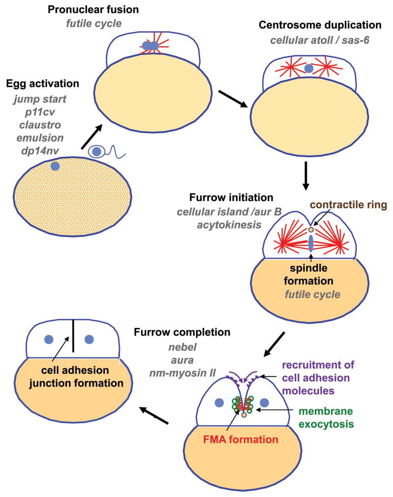
Diagram representing the main events associated with fertilization and cell division in the early zebrafish embryo. Maternal-effect mutants that affect various processes during this period are listed in grey type. See text for details.

Segregation of germ plasm components during cell division in the early zebrafish embryo. Top row of images: side views of embryos showing initially distinct localization of animal and vegetal germ plasm aggregates, and the animally-directed movement of the latter during the first cell cycles to the cells forming at the animal pole. Middle row of images: animal views of embryos. Animal germ plasm components are initially bound to a cortical filamentous network likely composed of f-actin (leftmost diagram). Peripheral movement of this network by the action of astral microtubules facilitates germ plasm aggregation and leads to the recruitment of germ plasm as a rod-like structure at the forming furrow (second and third diagram from left). Cytoskeletal rearrangements of the FMA are associated with further translocation and aggregation of the germ plasm at the distal ends of the mature furrow (fourth diagram from left). This process is repeated for the second furrow, resulting in four stable germ plasm aggregates (rightmost diagram). After their animally-directed movement, vegetal germ plasm components become anchored to the distal end of the compacted animal germ plasm, generating two associated subcompartments (rightmost diagrams in all rows). Bottom row: close up, in an animal view, of the cytoskeleton and associated germ plasm aggregates during the stages shown in the middle row diagrams. See text for additional details.
References
-
- Acilan C, Saunders WS. A tale of too many centrosomes. Cell. 2008;134:572–575. - PubMed
-
- Amatruda JF, Shepard JL, Stern HM, Zon LI. Zebrafish as a cancer model system. Cancer Cell. 2002;1:229–231. - PubMed
-
- Becker KA, Hart NH. The cortical actin cytoskeleton of unactivated zebrafish eggs: spatial organization and distribution of filamentous actin, non-filamentous actin, and myosin-II. Mol Reprod Dev. 1996;43:536–547. - PubMed
-
- Becker KA, Hart NH. Reorganization of filamentous actin and myosin-II in zebrafish eggs correlates temporally and spatially with cortical granule exocytosis. J Cell Sci. 1999;112:97–110. - PubMed
Publication types
MeSH terms
Substances
Grants and funding
LinkOut - more resources
Full Text Sources
