. 2010 Jul 1;127(1):127-37.
doi: 10.1002/ijc.25022.

Human BLCAP transcript: new editing events in normal and cancerous tissues

Affiliations
Free PMC article

Human BLCAP transcript: new editing events in normal and cancerous tissues

Federica Galeano et al. Int J Cancer. .
Free PMC article

Abstract

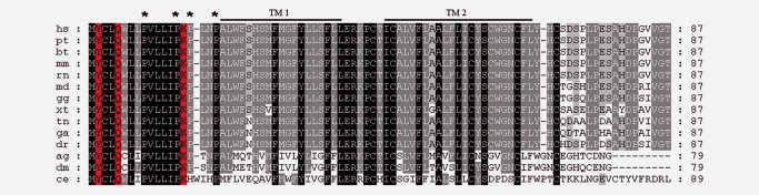
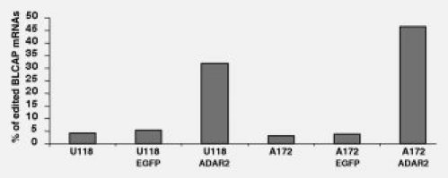
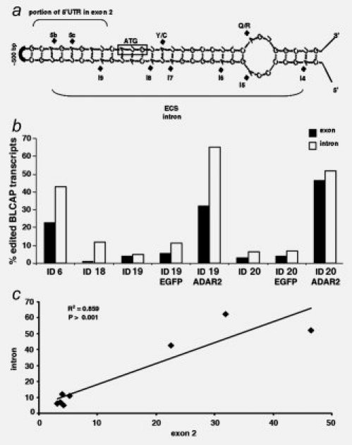
Bladder cancer-associated protein (BLCAP) is a highly conserved protein among species, and it is considered a novel candidate tumor suppressor gene originally identified from human bladder carcinoma. However, little is known about the regulation or the function of this protein. Here, we show that the human BLCAP transcript undergoes multiple A-to-I editing events. Some of the new editing events alter the highly conserved amino terminus of the protein creating alternative protein isoforms by changing the genetically coded amino acids. We found that both ADAR1 and ADAR2-editing enzymes cooperate to edit this transcript and that different tissues displayed distinctive ratios of edited and unedited BLCAP transcripts. Moreover, we observed a general decrease in BLCAP-editing level in astrocytomas, bladder cancer and colorectal cancer when compared with the related normal tissues. The newly identified editing events, found to be downregulated in cancers, could be useful for future studies as a diagnostic tool to distinguish malignancies or epigenetic changes in different tumors.


Figures

Figure 1
Figure 1
New editing events in the human BLCAP transcript. (a) Schematic representation of the BLCAP pre-mRNA. Exon 1 encoding the 5′UTR is represented in light blue with gray bars followed by the intron that divides the 5′UTR in 2. Exon 2 encodes the remaining 5′UTR (light blue with gray bars), the coding sequence (red rectangle) and the 3′UTR (light blue). Red dots indicate the editing sites in BLCAP mRNA (5′UTR and coding sequences). Red lines indicate the editing sites identified within the intron. (b) Sequences of BLCAP pre-mRNA. Adenosines in red indicate the edited sites in the intron (small letters) and exon 2 (capital letters). The underlined sequence corresponds to the coding region. The intron was found to be edited at 11 positions referred to as I1 to I11 from 5′ to 3′. The 5′UTR was edited at 3 positions referred to as: 5a, 5b and 5c. In the coding sequence, the edited positions are named according to the amino acid change they produce: Y/C, Q/R and K/R. Underneath, the sequences are shown the chromatograms of single clones after sequencing analysis of human BLCAP pre-mRNA in normal bladder tissue. As these sequences are cDNAs, the inosines are read as guanosines and the uridines as thymidines. The percentage of the editing events identified in the bladder tissue at different sites is shown in Table 3.
Figure 2
Figure 2
Multiple alignments of BLCAP proteins. Multiple alignments of BC10 proteins. Initials correspond to hs, Homo sapiens; pt, Pan troglodytes; bt, Bos taurus; mm, Mus musculus; rn, Rattus norvegicus; md, Monodelphis domestica; gg, Gallus gallus; xt, Xenopus tropicalis; tn, Tetraodon nigroviridis; ga, Gasterosteus aculeatus; dr, Danio rerio; ag, Anopheles gambiae; dm, Drosophila melanogaster; ce, Caenorhabditis elegans. Identical amino acid conservation among species is indicated in black. Residues identical in most of the species analyzed are indicated in gray. Indicated in red are the amino acids found to be edited. Black lines above the alignment indicate the hypothetical transmembrane domains (TM). Stars indicated the proline-rich motif at the N-terminus.
Figure 3
Figure 3
RNA editing in BLCAP mRNA. Schematic representation of human BLCAP mRNA. Exon 1 is depicted in bold. The remainder is exon 2 that encodes all the editing positions present in the mature BLCAP RNA, 3 within the 5′UTR and 3 within the coding sequence. The amino acid sequence is below the nucleotide sequence. The adenosines that are edited are bold and underlined. Arrows identify the position of the oligonucleotides hBLCAP5′F and hBLCAP3′RT used for the amplification of BLCAP after RT-PCR.
Figure 4
Figure 4
Increased editing activity observed on BLCAP mRNA. A172 and U118 astrocytoma cell lines were transfected with EGFP empty vector or a vector expressing EGFP-ADAR2. The editing events present in BLCAP mRNA are expressed as a percentage (y-axis). On the x-axis are the untransfected astrocytoma U118 and A172 cell lines, the cell lines transfected with EGFP vector and with EGFP-ADAR2. ADAR2 increased the number of BLCAP transcripts undergoing editing in both cell lines.
Figure 5
Figure 5
Alignment of BLCAP pre-mRNA in mammals. Alignment of BLCAP pre-mRNA among mammals identified by initials that correspond to hs, Homo sapiens; pt, Pan troglodytes; bt, Bos taurus; mm, Mus musculus; rn, Rattus norvegicus. (a) Upper alignment corresponds to the last ∼200 nucleotides of the BLCAP intron. (b) The lower alignment corresponds to the start of exon 2 in BLCAP. Nucleotide positions found edited in human are highlighted in green. The first ATG of the coding sequence corresponds to nucleotides 734–736 in (b). Sequences predicted to base pair to form an RNA duplex are highlighted in yellow. Identical nucleotides are indicated in black.
Figure 6
Figure 6
Comparison of editing within the intron and exon of BLCAP. (a) A portion of the predicted dsRNA structure formed between the intron and exon 2 is shown. The hypothetical ECS is also indicated. (b) The editing events present in BLCAP mRNA are expressed as a percentage (y-axis) and tissues, and cell lines are indicated with their ID identification number in all the samples studied (x-axis). The number of transcripts edited in the intron (in white bars) and in the exon (black bars) are shown. (c) Correlation of RNA editing between exon 2 and the intron in BLCAP. RNA editing within the BLCAP intron is expressed as a percentage (y-axis) and plotted against RNA editing within the BLCAP exon 2 (x-axis). Analysis was performed with linear regression; P values and R2 coefficients are shown.

References

    1. Bass BL. RNA editing by adenosine deaminases that act on RNA. Annu Rev Biochem. 2002;71:817–46. - PMC - PubMed
    1. Keegan LP, Gallo A, O'Connell MA. The many roles of an RNA editor. Nat Rev. 2001;2:869–78. - PubMed
    1. Basilio C, Wahba AJ, Lengyel P, Speyer JF, Ochoa S. Synthetic polynucleotides and the amino acid code. V. Proc Natl Acad Sci USA. 1962;48:613–6. - PMC - PubMed
    1. Rueter SM, Dawson TR, Emeson RB. Regulation of alternative splicing by RNA editing. Nature. 1999;399:75–80. - PubMed
    1. Nishikura K. Editor meets silencer: crosstalk between RNA editing and RNA interference. Nat Rev Mol Cell Biol. 2006;7:919–31. - PMC - PubMed

Publication types