

Notch signalling in ischaemia-induced angiogenesis
- PMID: 19909251
- PMCID: PMC2821013
- DOI: 10.1042/BST0371221
Notch signalling in ischaemia-induced angiogenesis
Abstract
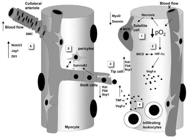
Notch signalling represents a key pathway essential for normal vascular development. Recently, great attention has been focused on the implication of Notch pathway components in postnatal angiogenesis and regenerative medicine. This paper critically reviews the most recent findings supporting the role of Notch in ischaemia-induced neovascularization. Notch signalling reportedly regulates several steps of the reparative process occurring in ischaemic tissues, including sprouting angiogenesis, vessel maturation, interaction of vascular cells with recruited leucocytes and skeletal myocyte regeneration. Further characterization of Notch interaction with other signalling pathways might help identify novel targets for therapeutic angiogenesis.
Figures
Similar articles
-
Inhibition of delta-like-4-mediated signaling impairs reparative angiogenesis after ischemia.Circ Res. 2010 Jul 23;107(2):283-93. doi: 10.1161/CIRCRESAHA.110.221663. Epub 2010 May 27. Circ Res. 2010. PMID: 20508179 Free PMC article.
-
Regulation of angiogenesis by homotypic and heterotypic notch signalling in endothelial cells and pericytes: from basic research to potential therapies.Angiogenesis. 2008;11(1):41-51. doi: 10.1007/s10456-008-9098-0. Epub 2008 Feb 7. Angiogenesis. 2008. PMID: 18256896 Review.
-
Notch signalling limits angiogenic cell behaviour in developing zebrafish arteries.Nature. 2007 Feb 15;445(7129):781-4. doi: 10.1038/nature05577. Epub 2007 Jan 28. Nature. 2007. PMID: 17259972
-
Notch1 promotes ordered revascularization through Semaphorin 3g modulation of downstream vascular patterning signalling factors.J Physiol. 2022 Feb;600(3):509-530. doi: 10.1113/JP282286. Epub 2022 Jan 17. J Physiol. 2022. PMID: 34921404 Free PMC article.
-
Role of Notch in endothelial biology.Angiogenesis. 2021 May;24(2):237-250. doi: 10.1007/s10456-021-09793-7. Epub 2021 May 29. Angiogenesis. 2021. PMID: 34050878 Review.
Cited by
-
Sustained release of naringin from silk-fibroin-nanohydroxyapatite scaffold for the enhancement of bone regeneration.Mater Today Bio. 2022 Jan 23;13:100206. doi: 10.1016/j.mtbio.2022.100206. eCollection 2022 Jan. Mater Today Bio. 2022. PMID: 35128373 Free PMC article.
-
Detrimental effect of class-selective histone deacetylase inhibitors during tissue regeneration following hindlimb ischemia.J Biol Chem. 2013 Aug 9;288(32):22915-29. doi: 10.1074/jbc.M113.484337. Epub 2013 Jul 7. J Biol Chem. 2013. PMID: 23836913 Free PMC article.
-
Notch signaling and cardiac repair.J Mol Cell Cardiol. 2012 Jun;52(6):1226-32. doi: 10.1016/j.yjmcc.2012.03.007. Epub 2012 Mar 21. J Mol Cell Cardiol. 2012. PMID: 22465038 Free PMC article. Review.
-
Effects of Transient Hypoxia versus Prolonged Hypoxia on Satellite Cell Proliferation and Differentiation In Vivo.Stem Cells Int. 2015;2015:961307. doi: 10.1155/2015/961307. Epub 2015 Feb 18. Stem Cells Int. 2015. PMID: 25788948 Free PMC article.
-
Dynamic Evaluation of Notch Signaling-Mediated Angiogenesis in Ischemic Rats Using Magnetic Resonance Imaging.Behav Neurol. 2018 May 6;2018:8351053. doi: 10.1155/2018/8351053. eCollection 2018. Behav Neurol. 2018. PMID: 29854019 Free PMC article.
References
-
- Selvin E, Erlinger TP. Prevalence of and risk factors for peripheral arterial disease in the United States: results from the National Health and Nutrition Examination Survey, 1999–2000. Circulation. 2004;110:738–743. - PubMed
-
- Slovut DP, Sullivan TM. Critical limb ischaemia: medical and surgical management. Vasc. Med. 2008;13:281–291. - PubMed
-
- Heilmann C, Beyersdorf F, Lutter G. Collateral growth: cells arrive at the construction site. Cardiovasc. Surg. 2002;10:570–578. - PubMed
-
- Emanueli C, Madeddu P. Therapeutic angiogenesis: translating experimental concepts to medically relevant goals. Vasc. Pharmacol. 2006;45:334–339. - PubMed
-
- Takeshita S, Pu LQ, Stein LA, Sniderman AD, Bunting S, Ferrara N, Isner JM, Symes JF. Intramuscular administration of vascular endothelial growth factor induces dose-dependent collateral artery augmentation in a rabbit model of chronic limb ischaemia. Circulation. 1994;90:II228–II234. - PubMed
Publication types
MeSH terms
Substances
Grants and funding
LinkOut - more resources
Full Text Sources