Review
. 2009 Nov;232(1):319-33.
doi: 10.1111/j.1600-065X.2009.00839.x.

SWAP-70-like adapter of T cells: a novel Lck-regulated guanine nucleotide exchange factor coordinating actin cytoskeleton reorganization and Ca2+ signaling in T cells

Affiliations
Review

SWAP-70-like adapter of T cells: a novel Lck-regulated guanine nucleotide exchange factor coordinating actin cytoskeleton reorganization and Ca2+ signaling in T cells

Stéphane Bécart et al. Immunol Rev. 2009 Nov.

Abstract

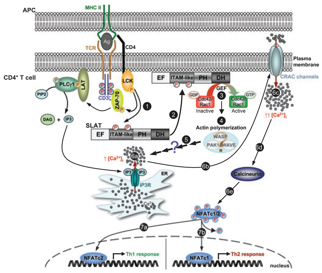
SWAP-70-like adapter of T cells (SLAT) is a recently identified guanine nucleotide exchange factor (GEF) for Cdc42 and Rac1, which is highly expressed in both thymocytes and peripheral T cells. Here, we present and discuss findings resulting from biochemical and genetic analyses aimed at unveiling the role of SLAT in CD4+ T-cell development, activation, and T-helper (Th) cell differentiation. Slat(-/-) mice display a developmental defect at one of the earliest stages of thymocyte differentiation, the double negative 1 (DN1) stage, leading to decreased peripheral T-cell numbers. Slat(-/-) peripheral CD4+ T cells demonstrate impaired T-cell receptor/CD28-induced proliferation and IL-2 production. Moreover, SLAT positively regulates the development of Th1 and Th2 inflammatory responses by controlling Ca2+/NFAT signaling. SLAT is also a positive regulator of the recently emerging Th subset, i.e., Th17 cells, as evidenced by its critical role in Th17 cell-mediated central nervous system inflammation. Furthermore, TCR engagement induces SLAT translocation to the immunological synapse, a process mediated by its Lck-dependent phosphorylation, which thereafter facilitates the triggering of SLAT GEF activity towards Cdc42 and Rac1, leading to NFAT activation and Th1/Th2 differentiation. Future work will aim to dissect the interacting partners of SLAT and may thus shed light on the poorly understood events that coordinate and link actin cytoskeleton reorganization to Ca2+ signaling and gene transcription in T cells.


Figures

Fig. 1
Fig. 1. Domain structure of SWAP-70-like adapter of T cells (SLAT)
Sequence analysis predicts the existence of a number of domains, which are shown in the figure. Numbers above the scheme refer to amino acid residues. A comparison of the ITAM-like sequence of SLAT with the consensus ITAM is also shown. Gray boxes indicate conserved amino acids in the ITAM. The ITAM-like domain contains negatively charged amino acids at conserved positions, the first YxxL/I motif, a spacer sequence of seven amino acids, and a second Tyr-containing sequence, YxxK, in which a lysine residue replaces the leucine/isoleucine typically found in ITAMs. The two tyrosines (Y) of this ITAM-like sequence, Tyr-133 and Tyr-144, are phosphorylated in a Lck-dependent manner upon TCR stimulation. The PH domain has the ability to bind PIP3 but is not required for TCR-induced translocation of SLAT to the membrane/IS. The DH domain has catalytic GEF activity towards Cdc42 and Rac1. The functionality and relevance of the putative EF hand domain and the nuclear localization signal (NLS) have yet to be determined.
Fig. 2
Fig. 2. A hypothetical model of SWAP-70-like adapter of T cells (SLAT) function in Ca2+ /NFAT signaling and Th cell differentiation
TCR stimulation leads to the sequential activation of protein tyrosine kinases such as Lck and ZAP-70, which phosphorylate adapter proteins such as LAT. Consequently, phosphorylated LAT binds the SH2 domain of PLCγ1, resulting in the phosphorylation/activation of PLCγ1. PLCγ1 catalyses the hydrolysis of the membrane phospholipid PIP2 to IP3 and DAG. IP3 binding to its receptor (IP3R) in the ER triggers Ca2+ release from intracellular stores, which, in return, triggers the opening of plasma membrane CRAC channels. Ca2+ influx through CRAC channels and elevated intracellular Ca2+ concentration activate Ca2+-dependent enzymes such as calcineurin, which dephosphorylates cytoplasmic NFATc1/2. Dephosphorylated NFATc1/2 translocate to the nucleus to induce gene transcription and influence Th cell responses; NFATc1 and NFATc2 positively regulate Th2 and Th1 responses, respectively. In this scenario, TCR engagement induces SLAT phosphorylation on Tyr-133 and Tyr-144 in its ITAM-like sequence (step 1), an event required for its translocation to the plasma membrane/IS (step 2). The exact mechanism mediating this relocalization remains elusive but does not involve the SLAT PH domain. Once at the membrane, additional TCR signals stimulate SLAT GEF activity towards Cdc42 and Rac1 (step 3), leading to activation of actin-regulatory proteins, e.g., WASP, WAVE, or PAK1, which promote actin polymerization (step 4). SLAT-mediated Cdc42/Rac1-dependent actin reorganization thereafter promotes Ca2+ release from intracellular stores (step 6a) and, consequently, Ca2+ influx (steps 6b-c) and NFAT activation (steps 6d-e), by acting downstream of IP3 production (step 5). The exact mechanism underlying this function is currently unknown. SLAT-mediated, Ca2+-dependent NFAT activation (steps 6a-c) controls Th1/Th2 commitment, depending upon the NFAT isoform involved (steps 7a and 7b, respectively).

References

    1. Aifantis I, Mandal M, Sawai K, Ferrando A, Vilimas T. Regulation of T-cell progenitor survival and cell-cycle entry by the pre-T-cell receptor. Immunol Rev. 2006;209:159–169. - PubMed
    1. Yamasaki S, Saito T. Molecular basis for pre-TCR-mediated autonomous signaling. Trends Immunol. 2007;28:39–43. - PubMed
    1. Jameson SC. Maintaining the norm: T-cell homeostasis. Nat Rev Immunol. 2002;2:547–556. - PubMed
    1. Freitas AA, Rocha B. Peripheral T cell survival. Curr Opin Immunol. 1999;11:152–156. - PubMed
    1. Mosmann TR, Coffman RL. TH1 and TH2 cells: different patterns of lymphokine secretion lead to different functional properties. Annu Rev Immunol. 1989;7:145–173. - PubMed

Publication types

MeSH terms

Substances