. 2010 Mar;138(3):969-80.e1-3.
doi: 10.1053/j.gastro.2009.11.004. Epub 2009 Nov 10.

Antimetastatic role of Smad4 signaling in colorectal cancer

Affiliations

Antimetastatic role of Smad4 signaling in colorectal cancer

Bixiang Zhang et al. Gastroenterology. 2010 Mar.

Abstract

Background & aims: Transforming growth factor (TGF)-beta signaling occurs through Smads 2/3/4, which translocate to the nucleus to regulate transcription; TGF-beta has tumor-suppressive effects in some tumor models and pro-metastatic effects in others. In patients with colorectal cancer (CRC), mutations or reduced levels of Smad4 have been correlated with reduced survival. However, the function of Smad signaling and the effects of TGF-beta-receptor kinase inhibitors have not been analyzed during CRC metastasis. We investigated the role of TGF-beta/Smad signaling in CRC progression.

Methods: We evaluated the role of TGF-beta/Smad signaling on cell proliferation, migration, invasion, tumorigenicity, and metastasis in Smad4-null colon carcinoma cell lines (MC38 and SW620) and in those that transgenically express Smad4. We also determined the effects of a TGF-beta-receptor kinase inhibitor (LY2109761) in CRC tumor progression and metastasis in mice.

Results: TGF-beta induced migration/invasion, tumorigenicity, and metastasis of Smad4-null MC38 and SW620 cells; incubation with LY2109761 reversed these effects. In mice, LY2109761 blocked metastasis of CRC cells to liver, inducing cancer cell expression of E-cadherin and reducing the expression of the tumorigenic proteins matrix metalloproteinase-9, nm23, urokinase plasminogen activator, and cyclooxygenase-2. Transgenic expression of Smad4 significantly reduced the oncogenic potential of MC38 and SW620 cells; in these transgenic cells, TGF-beta had tumor suppressor, rather than tumorigenic, effects.

Conclusions: TGF-beta/Smad signaling suppresses progression and metastasis of CRC cells and tumors in mice. Loss of Smad4 might underlie the functional shift of TGF-beta from a tumor suppressor to a tumor promoter; inhibitors of TGF-beta signaling might be developed as CRC therapeutics.


Figures

Figure 1
Figure 1
Smad-dependent signaling is inactivated in MC38 cells. (A) Lysates from TGF-β1 (5 ng/ml) treated MC38 and FET cells (positive control) were subjected to immunoprecipitation (IP) for Smad2 and Smad3 followed by western blotting with anti-Smad4 antibody (top panel). Lysates were subjected to western blot analyses with antibodies against indicated proteins. (B) MC38 cells were co-transfected with CMV-β-gal and p3TP-Lux or (CAGA)9 MLP-Luc with or without Smad4 expression construct. Cells were treated with TGF-β1 (5 ng/ml) and/or LY2109761 (5 μM). Luciferase activity was normalized to β-galactosidase activity and expressed as the mean ± S.D. of triplicate measurements. *P values <0.001 (For both reporters, the bars were compared as follows: third with first, fourth with third and sixth with fourth). (C) MC38 and SW620 cells were treated with TGF-β (5 ng/ml) for indicated time points, cell lysates were analyzed by western blotting with anti-ERK, phospho-ERK and -actin antibodies. Each experiment was repeated at least three times.
Figure 2
Figure 2
LY2109761 suppresses TGF-β-induced pro-oncogenic activities. (A) MC38 cells were treated with TGF-β (5 ng/ml) with LY2109761 (5 μM) for five days. Cells were counted every day and individual data points are presented as the mean ±S.D. of triplicate determinations. (B) MC38 cells that migrated through 8-μM pores in transwell plates in 5 h were fixed, stained and presented as the mean ± S.D. of three wells. MC38 cells were allowed to pass through the collagen layer (C) and the matrigel barrier (D) in transwell plates. Data are presented similarly as above. For figures 2B, 2C and 2D, *P value < 0.01 (Treatment with TGF-β and LY together compared to treatment with TGF-β alone) and **P values < 0.001 (Treatment with LY compared to treatment with TGF-β) (E) Wound healing assay was performed with MC38 cells as described in Materials and Methods. Original magnification, 200X. (F) MC38 cells were injected subcutaneously into the right flanks of C57BL/6 mice. LY2109761 or vehicle was administered orally twice a day. Tumors were measured and plotted as the mean volume ±S.D..
Figure 3
Figure 3
LY2109761 inhibits liver metastasis and prolongs survival. (A) Luciferase expressing-MC38 cells were injected into the spleens of C57BL/6 mice. Control mice (n=8) received vehicle and the treatment group was given (n=8) LY2109761 orally twice a day. Liver metastasis was monitored by bioluminescence imaging and images taken 23 days after splenic injection are presented. This experiment was repeated twice with similar results. (B) Integrated CT/microPET scan effectively shows reduction in liver metastasis following treatment with LY2109761. (C) Macroscopic liver metastases from control and LY2109761-treated mice are shown (top panel). LY2109761 reduced metastatic liver weight by more than 50% (bottom panel). *P values <0.001. (D) Lysates from livers of control and LY2109761-treated mice were analyzed with anti-phospho-Smad2 and anti-Smad2 antibodies. (E) Survival of mice treated with LY2109761 (n=6) or vehicle (n=6) was assayed after injecting MC38 cells into spleens of C57BL/6 mice. This experiment was repeated two times. *P<0.05
Figure 4
Figure 4
LY2109761 induces E-cadherin, and suppresses MMP-9, nm23, uPA and COX-2 and regulates cell cycle regulatory proteins. (A) & (B) Protein lysates from livers of control mice and LY2109761-treated mice were analyzed by western blotting as indicated. β-actin was used as loading control. NS: non-specific band. (C) Lysates from MC38 cells treated with LY2109761 (5 μM) were analyzed by western blotting with anti-E-cadherin antibodies.
Figure 5
Figure 5
Immunohistochemical analyses were performed using liver metastases of control and LY2109761-treated mice as described previously. Paraffin embedded blocks were prepared for serial sections and slides were processed for standard staining with H&E and Tunnel. Immunohistochemical analyses were performed with indicated antibodies. Original magnification, 630X.
Figure 6
Figure 6
Smad4 expression in MC38 cells re-establishes TGF-β-induced growth inhibition, and reduces migration and invasion. (A) Three stable Smad4 clones in MC38 cells were analyzed for Smad4 expression. (B) Immunoprecipitation assay was performed with anti-Smad2/Smad3 antibodies using lysates from TGF-β-treated MC38 cells/clones, followed by western blotting with anti-Smad4 antibody (top panel). Cell lysates were also analyzed for expression of Smad proteins as indicated. (C) For cell growth assay, cells from above clones were treated with 5 ng/ml TGF-β for five days and counted. Cell numbers in presence of TGF-β are presented considering the respective untreated cells as 100%. Individual data points are the mean ± S.D. of triplicate counts on fifth day. *P <0.01. Migration (D) and invasion (E) assays were performed with above MC38 stable Smad4 clones in presence or absence of TGF-β (5 ng/ml). Data points are the mean ±S.D. of migrated cells in three independent wells. For figures 6D and 6E, *P <0.01 (All three Smad4 clones were compared with the parental and vector control MC38 cells both in presence or absence of TGF-β).
Figure 7
Figure 7
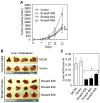
Stable re-expression of Smad4 reduces tumorigenicity and metastasis of MC38 cells. (A) Subcutaneous tumors were generated by the clones and tumor volumes were measured. Growth curves for tumors are presented as the mean ±S.D. of five tumors. *P<0.03, **P<0.05, ***P<0.01 (B) Liver metastasis was examined 21 days after injecting MC38 cells/clones into spleens of C57BL/6 mice as described above. (C) The graph shows average of the ratio between metastatic liver weight and total body weight of mice in each group (n=4). *P <0.001. This experiment was repeated two times.
Figure 8
Figure 8
Stable expression of Smad4 in SW620 cells reduces migration, invasion, tumorigenicity and metastasis. (A) Expression of Smad4 in stable clones generated from SW620 cells with CT26 cells as positive control. (B) Wound healing assay was performed using above clones as described in Materials and Methods. (C) Invasion assay using SW620 clones was done as described above. Data are presented as the mean ±S.D. from three independent wells. *P<0.001 (Smad4 clones with TGF-β treatment compared to parental and vector control cells with TGF-β treatment), **P<0.01. (Smad4 clones without TGF-β treatment compared to parental and vector control cells without TGF-β treatment) (D) In vivo tumorigenicity of SW620 clonal cells was assessed. The growth curves for tumors are presented as the mean ±S.D. of five tumors. * P<0.05, ** P<0.01 (Smad4 clones compared with controls). (E) SW620 clonal cells were injected into spleens of athymic nude mice (n=5, for parental cells n=7). Eight weeks after injection, mice were sacrificed and macroscopic liver metastasis or lymph node metastasis was assessed, as shown by circles in one mouse from each group. This experiment was repeated twice. (F) Data represent the effect of Smad4 re-expression on liver and lymph node metastasis. Number of mice with liver (including average number of foci per mouse) and lymph node metastases have been shown. A, P<0.001, B, P<0.002 compared to control by Student’s t-test.
Figure 8
Figure 8
Stable expression of Smad4 in SW620 cells reduces migration, invasion, tumorigenicity and metastasis. (A) Expression of Smad4 in stable clones generated from SW620 cells with CT26 cells as positive control. (B) Wound healing assay was performed using above clones as described in Materials and Methods. (C) Invasion assay using SW620 clones was done as described above. Data are presented as the mean ±S.D. from three independent wells. *P<0.001 (Smad4 clones with TGF-β treatment compared to parental and vector control cells with TGF-β treatment), **P<0.01. (Smad4 clones without TGF-β treatment compared to parental and vector control cells without TGF-β treatment) (D) In vivo tumorigenicity of SW620 clonal cells was assessed. The growth curves for tumors are presented as the mean ±S.D. of five tumors. * P<0.05, ** P<0.01 (Smad4 clones compared with controls). (E) SW620 clonal cells were injected into spleens of athymic nude mice (n=5, for parental cells n=7). Eight weeks after injection, mice were sacrificed and macroscopic liver metastasis or lymph node metastasis was assessed, as shown by circles in one mouse from each group. This experiment was repeated twice. (F) Data represent the effect of Smad4 re-expression on liver and lymph node metastasis. Number of mice with liver (including average number of foci per mouse) and lymph node metastases have been shown. A, P<0.001, B, P<0.002 compared to control by Student’s t-test.

Similar articles

Cited by

References

    1. Massagué J. TGF-β in Cancer. Cell. 2008;134:215–230. - PMC - PubMed
    1. Derynck R, Zhang YE. Smad-dependent and Smad-independent pathways in TGF-β family signalling. Nature. 2003;425:577–584. - PubMed
    1. Datta PK, Mann JR. Transforming growth factor-β (TGF- β) signling inhibitrs in cancer therapy. In: Jakowlew SB, editor. Transforming Growth Factor-β in Cancer Therapy. 1. Vol. 2. Totowa: Humana Press; 2007. pp. 573–588.
    1. Bandyopadhyay A, Agyin JK, Wang L, et al. Inhibition of pulmonary and skeletal metastasis by a transforming growth factor-β type I receptor kinase inhibitor. Cancer Res. 2006;66:6714–6721. - PubMed
    1. Ge R, Rajeev V, Ray P, et al. Inhibition of growth and metastasis of mouse mammary carcinoma by selective inhibitor of transforming growth factor-β type I receptor kinase in vivo. Clin Cancer Res. 2006;12:4315–4330. - PubMed

Publication types

MeSH terms