Review
. 2010 Mar 1;685(1-2):11-20.
doi: 10.1016/j.mrfmmm.2009.10.017. Epub 2009 Nov 10.

Uracil-DNA glycosylase: Structural, thermodynamic and kinetic aspects of lesion search and recognition

Affiliations
Review

Uracil-DNA glycosylase: Structural, thermodynamic and kinetic aspects of lesion search and recognition

Dmitry O Zharkov et al. Mutat Res. .

Abstract

Uracil appears in DNA as a result of cytosine deamination and by incorporation from the dUTP pool. As potentially mutagenic and deleterious for cell regulation, uracil must be removed from DNA. The major pathway of its repair is initiated by uracil-DNA glycosylases (UNG), ubiquitously found enzymes that hydrolyze the N-glycosidic bond of deoxyuridine in DNA. This review describes the current understanding of the mechanism of uracil search and recognition by UNG. The structure of UNG proteins from several species has been solved, revealing a specific uracil-binding pocket located in a DNA-binding groove. DNA in the complex with UNG is highly distorted to allow the extrahelical recognition of uracil. Thermodynamic studies suggest that UNG binds with appreciable affinity to any DNA, mainly due to the interactions with the charged backbone. The increase in the affinity for damaged DNA is insufficient to account for the exquisite specificity of UNG for uracil. This specificity is likely to result from multistep lesion recognition process, in which normal bases are rejected at one or several pre-excision stages of enzyme-substrate complex isomerization, and only uracil can proceed to enter the active site in a catalytically competent conformation. Search for the lesion by UNG involves random sliding along DNA alternating with dissociation-association events and partial eversion of undamaged bases for initial sampling.


Conflict of interest statement

Conflict of interest

This research was supported by the Presidium of the Russian Academy of Sciences and by Russian Foundation for Basic Research. The study sponsors were not involved in the study design, collection, analysis and interpretation of data, the writing of the manuscript, or the decision to submit the manuscript for publication.

Figures
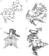
Fig. 1
Fig. 1
(A) schematic representation of hUNG structure. Circles indicate α-helices, triangles, β-sheets. The scheme is based on the structure of free hUNG (PDB ID 1AKZ [21]). (B) Structure of hUNG in a complex with DNA containing an uncleavable dUrd analog 2′-deoxypseudouridine (PDB ID 1EMH [25]). Protein is shown as ribbons, DNA, as sticks. (C) scheme of hUNG interactions with DNA after Ura excision (based on the structure of the enzyme–product complex, PDB ID 1SSP [24]). The residues are numbered relative to dUrd0, positive towards 5′, negative towards 3′; indices in the parentheses relate to the complementary strand. Only the central seven base pairs in the structure are shown. Hydrogen bonds are represented by arrows pointing to the acceptor; hydrophobic and van der Waals interactions are shown by direct contacts between the residues. Some hydrophobic/van der Waals contacts with unmodified residues are omitted for clarity. (D) Overlay of DNA structures from the non-specific/early recognition complex (PDB ID 2OXM [29]; light grey) and the specific/late recognition complex (PDB ID 1EMH [25]; dark grey). The void-filling Leu272 residue is also shown.
Fig. 2
Fig. 2
(A) Dependences of hUNG affinity for ss and ds ODNs of various composition on their length (n). (●) d(pA)n; (○) d(pA)n + d(pT)n; (■) d(pT)n; (◇) d(pC)n; (▲) d(pR)n (an oligomer consisting of n units of (3-hydroxytetrahydrofuran-2-yl)methyl phosphate, a deoxynucleotide analog lacking the nucleobase). The data are from Ref. [59]. (B) Determination of the electrostatic factor e reflecting an increase in the affinity due to one internucleotide phosphate group of d(pN)n using log dependence of factor f (an increase in the affinity for various d(pN)n per unit increase in length) on the relative hydrophobicity of mononucleotides. The relative hydrophobicities of mononucleotides were estimated by isocratic reverse-phase chromatography [98] and are expressed as the volume of retention on the column. Dependences for hUNG (■, e = 1.35), AP endonuclease APEX1 (●, e = 1.45) and DNA polymerase α (▲, e = 1.52) are shown; e factors are the intercepts of the linear dependences. The data are from Refs. [59,61,99].
Fig. 3
Fig. 3
Thermodynamic model for interaction of hUNG with non-specific d(pA)n:d(pT)n duplex (A) and specific dsDNA containing a dU deoxynucleotide (B). *Weak electrostatic interactions with internucleotide phosphate groups. Adenine bases forming weak hydrophobic interactions and/or van der Waals contacts with the enzyme are shadowed.
Fig. 4
Fig. 4
Oligonucleotide-based assay for studying processive cleavage by DNA glycosylases. The structure of the substrate is shown on the left. In the reaction scheme, P1 and EP1 refer to the substrate cleaved at any one of the Ura residues and to the respective enzyme–product complex; EP2, the complex with the substrate cleaved at one Ura and the enzyme translocated to the other Ura site; P2, the substrate cleaved at both Ura sites. Pcc, probability of correlated cleavage, i.e., the cleavage at the second Ura after the cleavage of the first Ura by the same enzyme molecule [82]. The initial rates of accumulation of 32-, 27-, and 19-mer products under the conditions of large substrate excess are denoted ν32, ν27, and ν19, respectively.

References

    1. Friedberg EC, Walker GC, Siede W, Wood RD, Schultz RA, Ellenberger T. DNA Repair and Mutagenesis. 3. ASM Press; Washington, DC: 2006.
    1. Lindahl T, Nyberg B. Heat-induced deamination of cytosine residues in deoxyribonucleic acid. Biochemistry. 1974;13:3405–3410. - PubMed
    1. Frederico LA, Kunkel TA, Shaw BR. A sensitive genetic assay for the detection of cytosine deamination: determination of rate constants and the activation energy. Biochemistry. 1990;29:2532–2537. - PubMed
    1. Tye BK, Chien J, Lehman IR, Duncan BK, Warner HR. Uracil incorporation: a source of pulse-labeled DNA fragments in the replication of the Escherichia coli chromosome. Proc Natl Acad Sci USA. 1978;75:233–237. - PMC - PubMed
    1. Lindahl T. An N-glycosidase from Escherichia coli that releases free uracil from DNA containing deaminated cytosine residues. Proc Natl Acad Sci USA. 1974;71:3649–3653. - PMC - PubMed

Publication types

LinkOut - more resources