Review
. 2010 Mar 8;62(3):284-304.
doi: 10.1016/j.addr.2009.11.002. Epub 2009 Nov 10.

Design and fabrication of magnetic nanoparticles for targeted drug delivery and imaging

Affiliations
Review

Design and fabrication of magnetic nanoparticles for targeted drug delivery and imaging

Omid Veiseh et al. Adv Drug Deliv Rev. .

Abstract

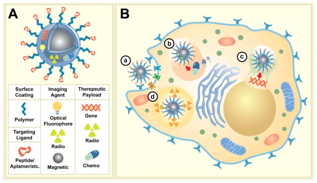
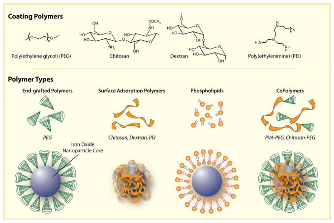
Magnetic nanoparticles (MNPs) represent a class of non-invasive imaging agents that have been developed for magnetic resonance (MR) imaging. These MNPs have traditionally been used for disease imaging via passive targeting, but recent advances have opened the door to cellular-specific targeting, drug delivery, and multi-modal imaging by these nanoparticles. As more elaborate MNPs are envisioned, adherence to proper design criteria (e.g. size, coating, molecular functionalization) becomes even more essential. This review summarizes the design parameters that affect MNP performance in vivo, including the physicochemical properties and nanoparticle surface modifications, such as MNP coating and targeting ligand functionalizations that can enhance MNP management of biological barriers. A careful review of the chemistries used to modify the surfaces of MNPs is also given, with attention paid to optimizing the activity of bound ligands while maintaining favorable physicochemical properties.


Figures

Figure 1
Figure 1
Illustrations with corresponding fluorescence images of ErbB2 receptor localization after treatment with different-sized heeceptin bound to gold NPs (Her–GNPs). In the fluorescence images of cells arrows indicate ErbB2 receptors, and the nucleus is counterstained with DAPI (blue) (scale bars=10 microm). Reprinted by permission from Macmillan Publishers Ltd: Nature Nanotechnology [102] Copyright 2008.
Figure 2
Figure 2
Conceptual scheme illustrating the varying multivalent affinity interactions between receptors on a cell surface and targeting ligands on a nanospheres versus a nanoworm. Conceptual adaptation from the figure previously published [71].
Figure 3
Figure 3
Illustration of multifunctional imaging/therapeutic MNPs anatomy and potential mechanisms of action at the cellular level. (A) A multifunctional MNP modified with targeting ligands extended from MNP surface with polymeric extenders, imaging reporters (optical, radio, magnetic), and potential therapeutic payloads (gene, radio, chemo). (B) Four possible modes of action for various therapeutic agents; a) Specific MNP binding to cell surface receptors (i.e. enzymes/proteins) facilitate their internalization and/or inactivation, b) controlled intercellular release of chemotherapeutics; c) release of gene therapeutic materials post endosomal escape and subsequent targeting of nucleus; and d) intracellular decay of radioactive materials.
Figure 4
Figure 4
Illustration depicting the assembly of polymers onto the surface of magnetic nanoparticle cores.
Figure 5
Figure 5
Illustration of the supermolecular assembly and presentation of targeting antibodies, proteins, peptides, aptamers and small molecules on the surfaces of SPIONs. Note that protein and antibody assembly is difficult to control. Small organic molecules do assemble well but their small size may cause their active targeting regions to be sterically blocked by polymeric coatings. Peptides and aptamers assembly can be controlled through their engineering, and can be modified to assemble in a manner that ensures their active sites are available for interaction with targets on cell surfaces.
Figure 6
Figure 6
TEM images showing increased membrane uptake subsequent to NP-CTX (NPC in figure) binding. Scale bars represent 5 mm for whole cell images (first row) and 200 nm for high magnification images (second row). White and black arrows identify NP-CTX and endosomes, respectively. Reproduced with permission from Wiley-VCH Verlag GmbH & Co. KGa: Small [159] Copyright 2009.
Figure 7
Figure 7
(a) In vivo MRI of mice bearing subcutaneous LS174T human colorectal adenocarcinoma (arrows). There was a significant drop in T2 relaxivity in images acquired after administration of the contrast agent (P = 0.003), indicating probe delivery. (b) A high-intensity NIRF signal on in vivo optical images associated with the tumor following injection of MNP-NIRF-siSurvivin confirmed the delivery of the probe to this tissue (left, white light; middle, NIRF; right, color-coded overlay). (c) Quantitative RT-PCR analysis of survivin expression in LS174T tumors after injection with either MNP-NIRF-siSurvivin, a mismatch control or the parental magnetic nanoparticle (MNP). Data are representative of three separate experiments. (d) Note distinct areas with a high density of apoptotic nuclei (green) in tumors treated with MNP-NIRF-siSurvivin (left). Such areas were not identified in tumors treated with the control MNPs (right). Sections were counter-stained with 6-diamidino-2-phenylindole (DAPI, blue). (e) H&E staining of frozen tumor sections revealed considerable eosinophilic areas of tumor necrosis (N) in tumors treated with MNP-NIRF-siSurvivin (left). Tumors treated with MNPs were devoid of necrotic tissue (right). Purple hematoxiphilic regions (V) indicate viable tumor tissues. Scale bar, 50 μm. Reprinted by permission from Macmillan Publishers Ltd: Nature Medicine [201], copyright 2007.
Figure 8
Figure 8
Axial cross sections displaying 9L tumors of mice before injection of nanoparticle conjugates and 1 and 3 days post-injection. T2 map overlays of the tumor region show decreased T2 for both NP-MTX and NP-MTX-CTX nanoprobe conjugates 1 day after administration. However, the reduction is more significant and uniform in tumor of mouse receiving NP-MTX-CTX. A total of 3 days post-injection, the tumor T2 values of the mouse receiving NP-MTX-CTX remained at the decreased level, while those of mouse receiving NP-MTX returned to the post-injection level suggesting clearance of NP-MTX from tumor tissue. (CTX = Chlorotoxin, MTX = Methotrexate, NP = Nanoparticle). Reproduced with permission from Future Medicine Ltd: Nanomedicine [156] Copyright 2008.
Figure 9
Figure 9
a) MR images and their color maps (tumor region) of cancer-targeting events of HER-MMPNs (a-d) and IRR-MMPNs (e-h) in NIH3T6.7 cells implanted in mice at various time intervals: a,e) preinjection; b,f) immediately; c,g) 1 h; d,h) 12 h after injection of the MMPNs. i) ΔR2/R2pre graph versus time before and after injection of MMPNs. j) Comparative therapeutic-efficacy study in an in vivo model. Reproduced with permission from Wiley-VCH Verlag GmbH & Co. KGa: Angew. Chem. Int. Ed. [241]

Similar articles

Cited by

References

    1. Ferrari M. Cancer nanotechnology: Opportunities and challenges. Nature Reviews Cancer. 2005;5:161–171. - PubMed
    1. Sanvicens N, Marco MP. Multifunctional nanoparticles - properties and prospects for their use in human medicine. Trends in Biotechnology. 2008;26:425–433. - PubMed
    1. Farokhzad OC, Langer R. Nanomedicine: Developing smarter therapeutic and diagnostic modalities. Advanced Drug Delivery Reviews. 2006;58:1456–1459. - PubMed
    1. He J, VanBrocklin HF, Franc BL, Seo Y, Jones EF. Nanoprobes for medical diagnosis: Current status of nanotechnology in molecular imaging. Current Nanoscience. 2008;4:17–29.
    1. Matsuura N, Rowlands JA. Towards new functional nanostructures for medical imaging. Medical Physics. 2008;35:4474–4487. - PubMed

Publication types