

Cathepsin G: roles in antigen presentation and beyond
- PMID: 19910052
- PMCID: PMC4159238
- DOI: 10.1016/j.molimm.2009.10.003
Cathepsin G: roles in antigen presentation and beyond
Abstract
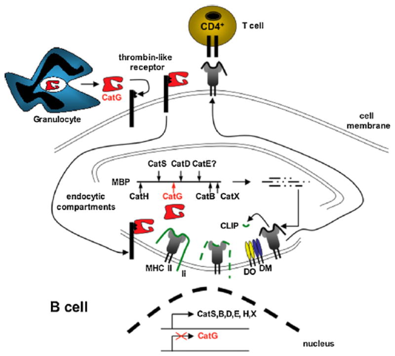
Contributions from multiple cathepsins within endosomal antigen processing compartments are necessary to process antigenic proteins into antigenic peptides. Cysteine and aspartyl cathepsins have been known to digest antigenic proteins. A role for the serine protease, cathepsin G (CatG), in this process has been described only recently, although CatG has long been known to be a granule-associated proteolytic enzyme of neutrophils. In line with a role for this enzyme in antigen presentation, CatG is found in endocytic compartments of a variety of antigen presenting cells. CatG is found in primary human monocytes, B cells, myeloid dendritic cells 1 (mDC1), mDC2, plasmacytoid DC (pDC), and murine microglia, but is not expressed in B cell lines or monocyte-derived DC. Purified CatG can be internalized into endocytic compartments in CatG non-expressing cells, widening the range of cells where this enzyme may play a role in antigen processing. Functional assays have implicated CatG as a critical enzyme in processing of several antigens and autoantigens. In this review, historical and recent data on CatG expression, distribution, function and involvement in disease will be summarized and discussed, with a focus on its role in antigen presentation and immune-related events.
Copyright 2010 Elsevier Ltd. All rights reserved.
Figures
Similar articles
-
Cathepsin G is differentially expressed in primary human antigen-presenting cells.Cell Immunol. 2009;255(1-2):41-5. doi: 10.1016/j.cellimm.2008.10.001. Epub 2008 Nov 25. Cell Immunol. 2009. PMID: 19036358
-
Masking of a cathepsin G cleavage site in vivo contributes to the proteolytic resistance of major histocompatibility complex class II molecules.Immunology. 2010 Jul;130(3):436-46. doi: 10.1111/j.1365-2567.2010.03247.x. Epub 2010 Mar 17. Immunology. 2010. PMID: 20331476 Free PMC article.
-
Invariant chain processing is independent of cathepsin variation between primary human B cells/dendritic cells and B-lymphoblastoid cells.Cell Immunol. 2011;269(2):96-103. doi: 10.1016/j.cellimm.2011.03.012. Epub 2011 Mar 17. Cell Immunol. 2011. PMID: 21543057
-
Cathepsin G and its Dichotomous Role in Modulating Levels of MHC Class I Molecules.Arch Immunol Ther Exp (Warsz). 2020 Aug 19;68(4):25. doi: 10.1007/s00005-020-00585-3. Arch Immunol Ther Exp (Warsz). 2020. PMID: 32815043 Review.
-
The lysosomal cysteine proteases in MHC class II antigen presentation.Immunol Rev. 2005 Oct;207:229-41. doi: 10.1111/j.0105-2896.2005.00310.x. Immunol Rev. 2005. PMID: 16181340 Review.
Cited by
-
Cathepsin G induces cell aggregation of human breast cancer MCF-7 cells via a 2-step mechanism: catalytic site-independent binding to the cell surface and enzymatic activity-dependent induction of the cell aggregation.Mediators Inflamm. 2012;2012:456462. doi: 10.1155/2012/456462. Epub 2012 Jul 8. Mediators Inflamm. 2012. PMID: 22919124 Free PMC article.
-
The Upregulation of Cathepsin G Is Associated with Resistance to Bovine Paratuberculosis.Animals (Basel). 2022 Nov 4;12(21):3038. doi: 10.3390/ani12213038. Animals (Basel). 2022. PMID: 36359162 Free PMC article.
-
ABPP and Host-Virus Interactions.Curr Top Microbiol Immunol. 2019;420:131-154. doi: 10.1007/82_2018_139. Curr Top Microbiol Immunol. 2019. PMID: 30244323 Free PMC article. Review.
-
A Local Inflammatory Renin-Angiotensin System Drives Sensory Axon Sprouting in Provoked Vestibulodynia.J Pain. 2017 May;18(5):511-525. doi: 10.1016/j.jpain.2016.12.008. Epub 2017 Jan 3. J Pain. 2017. PMID: 28062309 Free PMC article.
-
The Intricate Balance between Life and Death: ROS, Cathepsins, and Their Interplay in Cell Death and Autophagy.Int J Mol Sci. 2024 Apr 6;25(7):4087. doi: 10.3390/ijms25074087. Int J Mol Sci. 2024. PMID: 38612897 Free PMC article. Review.
References
-
- Allen DH, Tracy PB. Human coagulation factor V is activated to the functional cofactor by elastase and cathepsin G expressed at the monocyte surface. J Biol Chem. 1995;270:1408–15. - PubMed
-
- Anderssen T, Halvorsen H, Bajaj SP, Osterud B. Human leukocyte elastase and cathepsin G inactivate factor VII by limited proteolysis. Thromb Haemost. 1993;70:414–7. - PubMed
-
- Baggiolini M, Bretz U, Dewald B, Feigenson ME. The polymorphonuclear leukocyte. Agents Actions. 1978;8:3–10. - PubMed
-
- Baggiolini M, Schnyder J, Bretz U, Dewald B, Ruch W. Cellular mechanisms of proteinase release from inflammatory cells and the degradation of extracellular proteins. Ciba Found Symp. 1979:105–21. - PubMed
Publication types
MeSH terms
Substances
Grants and funding
LinkOut - more resources
Full Text Sources
Other Literature Sources
Research Materials
Miscellaneous
