

Mitochondrial-targeted fluorescent probes for reactive oxygen species
- PMID: 19910238
- PMCID: PMC2830890
- DOI: 10.1016/j.cbpa.2009.10.014
Mitochondrial-targeted fluorescent probes for reactive oxygen species
Abstract
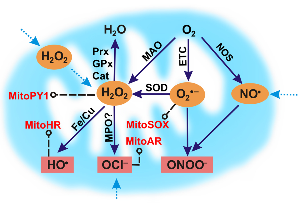
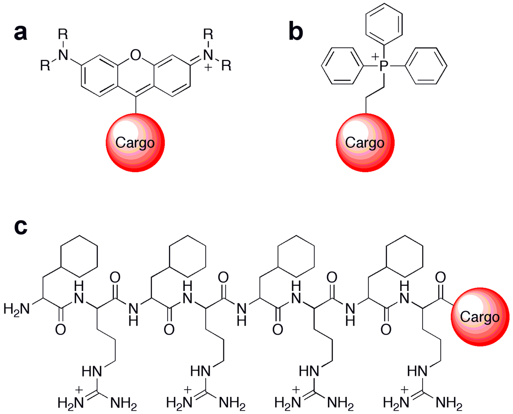
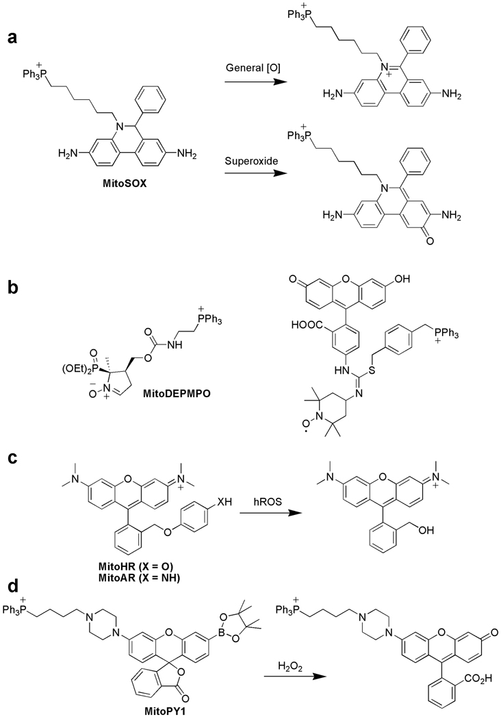
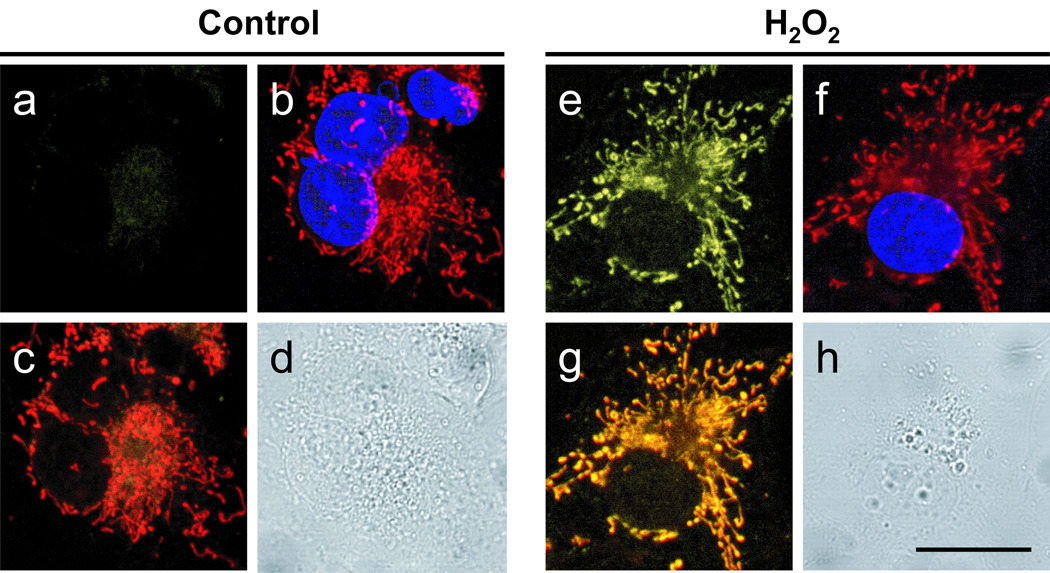
As the primary consumers of oxygen within all aerobic organisms, mitochondria are a major source of cellular reactive oxygen species (ROS) derived from the in vivo chemistry of oxygen metabolism. Mitochondrial ROS have been traditionally implicated in aging and in a variety of pathologies, including cancer, neurodegeneration, and diabetes, but recent studies also link controlled mitochondrial ROS fluxes to cell regulation and signaling events. Progress in the development of mitochondrial-targeted fluorescent small-molecule indicators that detect specific ROS with high selectivity offers a promising approach for interrogating mitochondrial ROS production, trafficking, and downstream biological effects.
2009 Elsevier Ltd. All rights reserved.
Figures
Similar articles
-
Cytometric assessment of mitochondria using fluorescent probes.Cytometry A. 2011 Jun;79(6):405-25. doi: 10.1002/cyto.a.21061. Cytometry A. 2011. PMID: 21595013 Review.
-
Imaging mitochondrial reactive oxygen species with fluorescent probes: current applications and challenges.Free Radic Res. 2015 Apr;49(4):374-82. doi: 10.3109/10715762.2015.1014813. Epub 2015 Mar 19. Free Radic Res. 2015. PMID: 25789762 Review.
-
Fluorescent probes for sensing and imaging biological hydrogen sulfide.Curr Opin Chem Biol. 2012 Dec;16(5-6):595-601. doi: 10.1016/j.cbpa.2012.07.014. Epub 2012 Aug 23. Curr Opin Chem Biol. 2012. PMID: 22921406 Free PMC article. Review.
-
Sub-organellar mitochondrial hydrogen peroxide observed using a SNAP tag targeted coumarin-based fluorescent reporter.Redox Biol. 2025 Mar;80:103502. doi: 10.1016/j.redox.2025.103502. Epub 2025 Jan 20. Redox Biol. 2025. PMID: 39864323 Free PMC article.
-
In vivo ROS and redox potential fluorescent detection in plants: Present approaches and future perspectives.Methods. 2016 Oct 15;109:92-104. doi: 10.1016/j.ymeth.2016.07.009. Epub 2016 Jul 14. Methods. 2016. PMID: 27424086 Review.
Cited by
-
Preparation and use of MitoPY1 for imaging hydrogen peroxide in mitochondria of live cells.Nat Protoc. 2013 Jun;8(6):1249-59. doi: 10.1038/nprot.2013.064. Epub 2013 May 30. Nat Protoc. 2013. PMID: 23722262 Free PMC article.
-
Nutritional countermeasures targeting reactive oxygen species in cancer: from mechanisms to biomarkers and clinical evidence.Antioxid Redox Signal. 2013 Dec 10;19(17):2157-96. doi: 10.1089/ars.2012.4662. Epub 2013 Apr 15. Antioxid Redox Signal. 2013. PMID: 23458328 Free PMC article. Review.
-
A novel highly specific and ultrasensitive fluorescent probe for monitoring hypochlorous acid and its application in live cells.RSC Adv. 2019 Feb 6;9(8):4659-4664. doi: 10.1039/c8ra09551e. eCollection 2019 Jan 30. RSC Adv. 2019. PMID: 35520202 Free PMC article.
-
Simultaneous fluorescence imaging of hydrogen peroxide in mitochondria and endoplasmic reticulum during apoptosis.Chem Sci. 2016 Sep 1;7(9):6153-6159. doi: 10.1039/c6sc01793b. Epub 2016 Jun 1. Chem Sci. 2016. PMID: 30034754 Free PMC article.
-
A unique approach toward near-infrared fluorescent probes for bioimaging with remarkably enhanced contrast.Chem Sci. 2016 Mar 1;7(3):2275-2285. doi: 10.1039/c5sc04014k. Epub 2016 Jan 4. Chem Sci. 2016. PMID: 29910917 Free PMC article.
References
-
- Stone JR, Yang S. Hydrogen peroxide: a signaling messenger. Antioxid Redox Sign. 2006;8:243–270. - PubMed
-
- Rhee SG. Cell signaling: H2O2, a necessary evil for cell signaling. Science. 2006;312:1882–1883. - PubMed
-
- Veal EA, Day AM, Morgan BA. Hydrogen peroxide sensing and signaling. Mol Cell. 2007;26:1–14. - PubMed
-
- D'AutrEaux B, Toledano MB. ROS as signalling molecules: mechanisms that generate specificity in ROS homeostasis. Nat Mol Cell Biol. 2007;8:813–824. - PubMed
-
- Giorgio M, Trinei M, Migliaccio E, Pelicci PG. Hydrogen peroxide: a metabolic by-product or a common mediator of ageing signals? Nat Mol Cell Biol. 2007;8:722–728. - PubMed
Publication types
MeSH terms
Substances
Grants and funding
LinkOut - more resources
Full Text Sources
Other Literature Sources
Research Materials
