

Partial seizures are associated with early increases in signal complexity
- PMID: 19910249
- PMCID: PMC2818227
- DOI: 10.1016/j.clinph.2009.09.018
Partial seizures are associated with early increases in signal complexity
Abstract
Objectives: Partial seizures are often believed to be associated with EEG signals of low complexity because seizures are associated with increased neural network synchrony. The investigations reported here provide an assessment of the signal complexity of epileptic seizure onsets using newly developed quantitative measures.
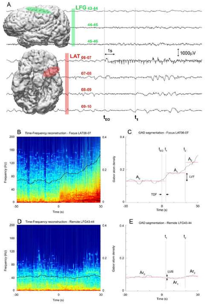
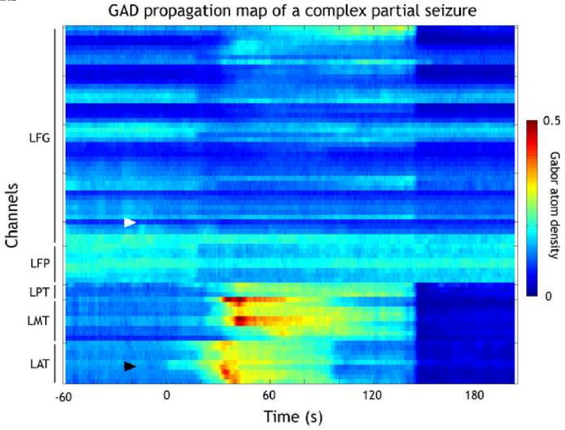
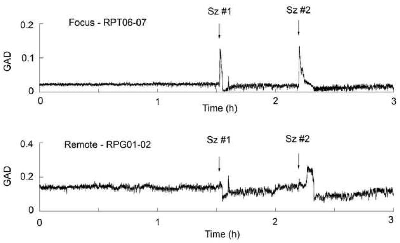
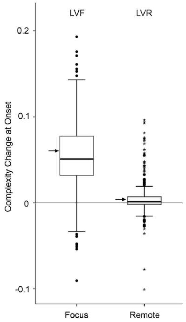
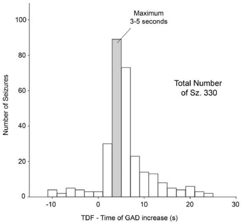
Methods: Using the Gabor atom density (GAD) measure of signal complexity, 339 partial seizures in 45 patients with intracranial electrode arrays were analyzed. Segmentation procedures were applied to determine the timing and amplitude of GAD changes relative to the electrographic onset of the seizure.
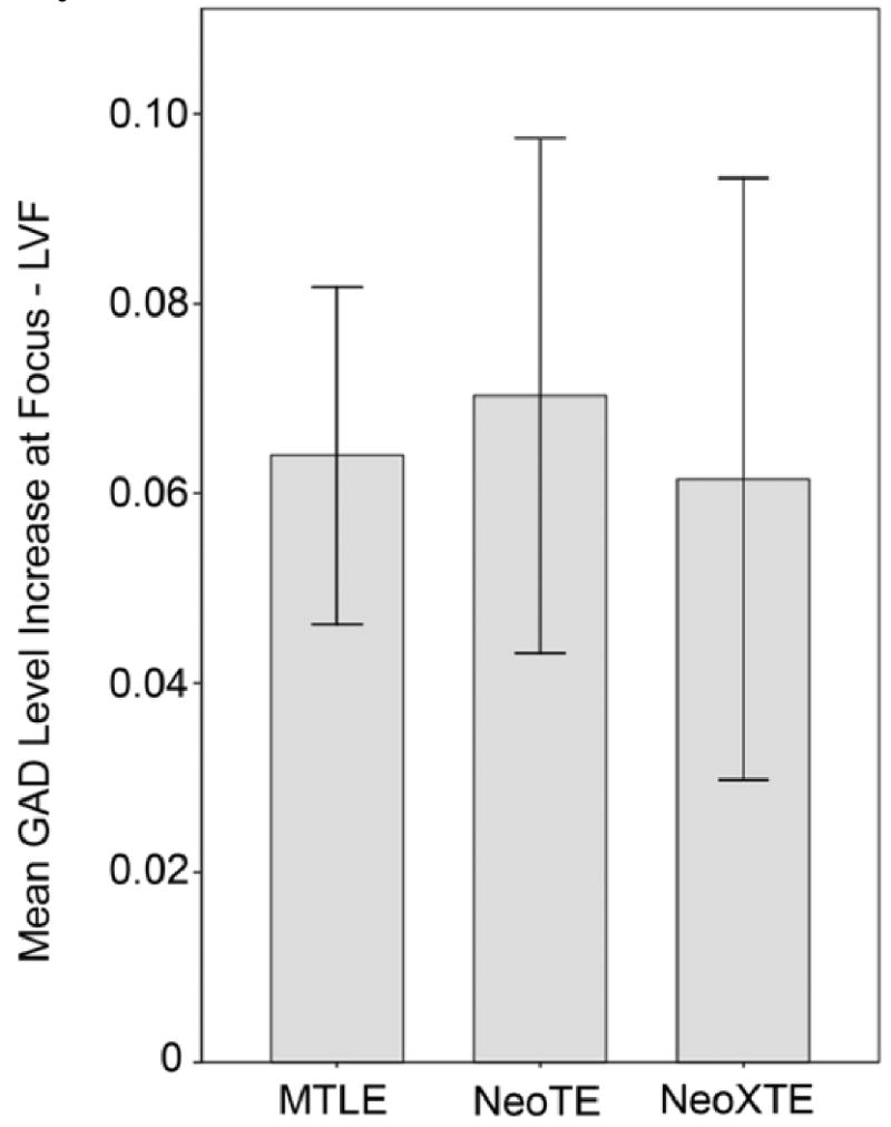
Results: Three hundred and thirty out of 339 seizures have significant complexity level changes, with 319 (97%) having an increase in complexity. GAD increases occur within seconds of the onset of the partial seizure but are not observed in channels remote from the focus. The complexity increase is similar for seizures from mesial temporal origin, neocortical temporal and extra-temporal origin.
Conclusions: Partial onset seizures are associated with early increases in signal complexity as measured by GAD. This increase is independent of the location of the seizure focus.
Significance: Despite the often predominant rhythmic activity that characterizes onset and early evolution of epileptic seizures, partial seizure onset is associated with an early increase in complexity. These changes are common to partial seizures originating from different brain regions, indicating a similar seizure dynamic.
Copyright 2009 International Federation of Clinical Neurophysiology. Published by Elsevier Ireland Ltd. All rights reserved.
Figures
Similar articles
-
Characterization of early partial seizure onset: frequency, complexity and entropy.Clin Neurophysiol. 2012 Apr;123(4):658-69. doi: 10.1016/j.clinph.2011.08.003. Epub 2011 Aug 26. Clin Neurophysiol. 2012. PMID: 21872526 Free PMC article.
-
Intrinsic ictal dynamics at the seizure focus: effects of secondary generalization revealed by complexity measures.Epilepsia. 2007 Feb;48(2):297-304. doi: 10.1111/j.1528-1167.2006.00963.x. Epilepsia. 2007. PMID: 17295623
-
Impaired consciousness in temporal lobe seizures: role of cortical slow activity.Brain. 2010 Dec;133(Pt 12):3764-77. doi: 10.1093/brain/awq316. Epub 2010 Nov 16. Brain. 2010. PMID: 21081551 Free PMC article.
-
Ictal vomiting as a sign of temporal lobe epilepsy confirmed by stereo-EEG and surgical outcome.Epilepsy Behav. 2015 Dec;53:112-6. doi: 10.1016/j.yebeh.2015.10.009. Epub 2015 Nov 8. Epilepsy Behav. 2015. PMID: 26558713 Review.
-
Hypothalamic hamartoma: Epileptogenesis beyond the lesion?Epilepsia. 2017 Jun;58 Suppl 2:32-40. doi: 10.1111/epi.13755. Epilepsia. 2017. PMID: 28591482 Review.
Cited by
-
Characterization of early partial seizure onset: frequency, complexity and entropy.Clin Neurophysiol. 2012 Apr;123(4):658-69. doi: 10.1016/j.clinph.2011.08.003. Epub 2011 Aug 26. Clin Neurophysiol. 2012. PMID: 21872526 Free PMC article.
-
Information dynamics of in silico EEG Brain Waves: Insights into oscillations and functions.PLoS Comput Biol. 2024 Sep 5;20(9):e1012369. doi: 10.1371/journal.pcbi.1012369. eCollection 2024 Sep. PLoS Comput Biol. 2024. PMID: 39236071 Free PMC article.
-
Controversies in epilepsy: debates held during the Fourth International Workshop on Seizure Prediction.Epilepsy Behav. 2010 Sep;19(1):4-16. doi: 10.1016/j.yebeh.2010.06.009. Epub 2010 Aug 13. Epilepsy Behav. 2010. PMID: 20708976 Free PMC article.
-
Preictal dynamics of EEG complexity in intracranially recorded epileptic seizure: a case report.Medicine (Baltimore). 2014 Nov;93(23):e151. doi: 10.1097/MD.0000000000000151. Medicine (Baltimore). 2014. PMID: 25415671 Free PMC article.
-
Navigated Transcranial Magnetic Stimulation (nTMS) based Preoperative Planning for Brain Tumor Treatment.CNS Neurol Disord Drug Targets. 2024;23(7):883-893. doi: 10.2174/1871527322666230619103429. CNS Neurol Disord Drug Targets. 2024. PMID: 37340739 Review.
References
-
- Afra P, Jouny CC, Bergey GK. Duration of complex partial seizures: an intracranial EEG study. Epilepsia. 2008;49:677–684. - PubMed
-
- Alarcon G, Binnie CD, Elwes RD, Polkey CE. Power spectrum and intracranial EEG patterns at seizure onset in partial epilepsy. Electroencephalogr Clin Neurophysiol. 1995;94:326–337. - PubMed
-
- Babloyantz A, Salazar J, Nicolis C. Evidence of chaotic dynamics of brain activity during the sleep cycle. Phys Lett A. 1985;111:152–156.
-
- Bergey GK, Franaszczuk PJ. Epileptic seizures are characterized by changing signal complexity. Clin Neurophysiol. 2001;112:241–249. - PubMed
Publication types
MeSH terms
Grants and funding
LinkOut - more resources
Full Text Sources
