

MeMotif: a database of linear motifs in alpha-helical transmembrane proteins
- PMID: 19910368
- PMCID: PMC2808916
- DOI: 10.1093/nar/gkp1042
MeMotif: a database of linear motifs in alpha-helical transmembrane proteins
Abstract
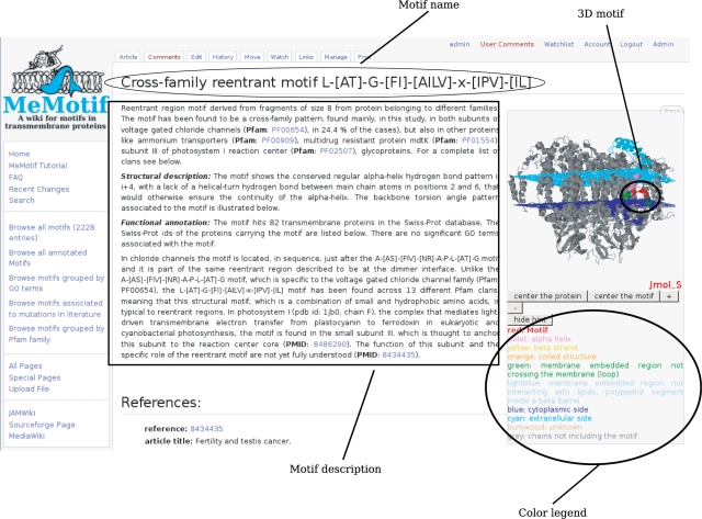
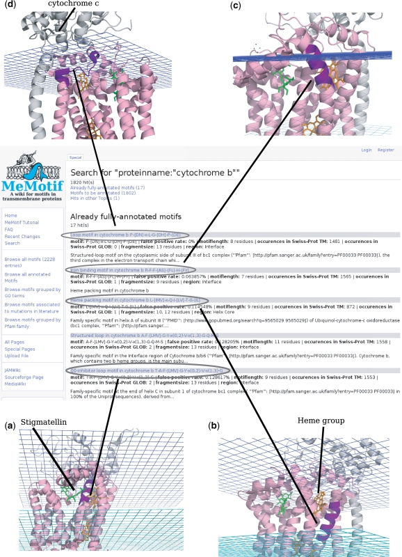
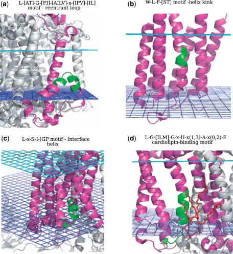
Membrane proteins are important for many processes in the cell and used as main drug targets. The increasing number of high-resolution structures available makes for the first time a characterization of local structural and functional motifs in alpha-helical transmembrane proteins possible. MeMotif (http://projects.biotec.tu-dresden.de/memotif) is a database and wiki which collects more than 2000 known and novel computationally predicted linear motifs in alpha-helical transmembrane proteins. Motifs are fully described in terms of several structural and functional features and editable. Motifs contained in MeMotif can be used in different biological applications, from the identification of biochemically important functional residues which are candidates for mutagenesis experiments to the improvement of tools for transmembrane protein modeling.
Figures
Similar articles
-
ELM: the status of the 2010 eukaryotic linear motif resource.Nucleic Acids Res. 2010 Jan;38(Database issue):D167-80. doi: 10.1093/nar/gkp1016. Epub 2009 Nov 17. Nucleic Acids Res. 2010. PMID: 19920119 Free PMC article.
-
Protein Geometry Database: a flexible engine to explore backbone conformations and their relationships to covalent geometry.Nucleic Acids Res. 2010 Jan;38(Database issue):D320-5. doi: 10.1093/nar/gkp1013. Epub 2009 Nov 11. Nucleic Acids Res. 2010. PMID: 19906726 Free PMC article.
-
CyanoBase: the cyanobacteria genome database update 2010.Nucleic Acids Res. 2010 Jan;38(Database issue):D379-81. doi: 10.1093/nar/gkp915. Epub 2009 Oct 30. Nucleic Acids Res. 2010. PMID: 19880388 Free PMC article.
-
Identification of motifs in protein sequences.Curr Protoc Cell Biol. 2001 May;Appendix 1:Appendix 1C. doi: 10.1002/0471143030.cba01cs00. Curr Protoc Cell Biol. 2001. PMID: 18228275 Review.
-
From interactions of single transmembrane helices to folding of alpha-helical membrane proteins: analyzing transmembrane helix-helix interactions in bacteria.Curr Protein Pept Sci. 2007 Feb;8(1):45-61. doi: 10.2174/138920307779941578. Curr Protein Pept Sci. 2007. PMID: 17305560 Review.
Cited by
-
Deorphanizing the human transmembrane genome: A landscape of uncharacterized membrane proteins.Acta Pharmacol Sin. 2014 Jan;35(1):11-23. doi: 10.1038/aps.2013.142. Epub 2013 Nov 18. Acta Pharmacol Sin. 2014. PMID: 24241348 Free PMC article. Review.
-
Expediting topology data gathering for the TOPDB database.Nucleic Acids Res. 2015 Jan;43(Database issue):D283-9. doi: 10.1093/nar/gku1119. Epub 2014 Nov 11. Nucleic Acids Res. 2015. PMID: 25392424 Free PMC article.
-
Helix kinks are equally prevalent in soluble and membrane proteins.Proteins. 2014 Sep;82(9):1960-70. doi: 10.1002/prot.24550. Epub 2014 Apr 16. Proteins. 2014. PMID: 24638929 Free PMC article.
-
Transferring functional annotations of membrane transporters on the basis of sequence similarity and sequence motifs.BMC Bioinformatics. 2013 Nov 28;14:343. doi: 10.1186/1471-2105-14-343. BMC Bioinformatics. 2013. PMID: 24283849 Free PMC article.
-
Computational modeling of membrane proteins.Proteins. 2015 Jan;83(1):1-24. doi: 10.1002/prot.24703. Epub 2014 Nov 19. Proteins. 2015. PMID: 25355688 Free PMC article. Review.
