. 2010 Feb;156(Pt 2):356-373.
doi: 10.1099/mic.0.033241-0. Epub 2009 Nov 12.

Role of host cell polarity and leading edge properties in Pseudomonas type III secretion

Affiliations

Role of host cell polarity and leading edge properties in Pseudomonas type III secretion

Dacie R Bridge et al. Microbiology (Reading). 2010 Feb.

Abstract

Type III secretion (T3S) functions in establishing infections in a large number of Gram-negative bacteria, yet little is known about how host cell properties might function in this process. We used the opportunistic pathogen Pseudomonas aeruginosa and the ability to alter host cell sensitivity to Pseudomonas T3S to explore this problem. HT-29 epithelial cells were used to study cellular changes associated with loss of T3S sensitivity, which could be induced by treatment with methyl-beta-cyclodextrin or perfringolysin O. HL-60 promyelocytic cells are innately resistant to Pseudomonas T3S and were used to study cellular changes occurring in response to induction of T3S sensitivity, which occurred following treatment with phorbol esters. Using both cell models, a positive correlation was observed between eukaryotic cell adherence to tissue culture wells and T3S sensitivity. In examining the type of adhesion process linked to T3S sensitivity in HT-29 cells, a hierarchical order of protein involvement was identified that paralleled the architecture of leading edge (LE) focal complexes. Conversely, in HL-60 cells, induction of T3S sensitivity coincided with the onset of LE properties and the development of actin-rich projections associated with polarized cell migration. When LE architecture was examined by immunofluorescent staining for actin, Rac1, IQ-motif-containing GTPase-activating protein 1 (IQGAP1) and phosphatidylinositol 3 kinase (PI3 kinase), intact LE structure was found to closely correlate with host cell sensitivity to P. aeruginosa T3S. Our model for host cell involvement in Pseudomonas T3S proposes that cortical actin polymerization at the LE alters membrane properties to favour T3S translocon function and the establishment of infections, which is consistent with Pseudomonas infections targeting wounded epithelial barriers undergoing cell migration.


Figures

Fig. 1.
Fig. 1.
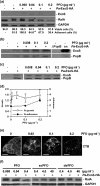
Effect of MβCD treatment on Pa-T3S sensitivity. HT-29 cells were treated with the indicated concentration of MβCD for 30 min, prior to and during co-culture with strain Pa-ExoS-HA. (a) To assay for Pa-T3S sensitivity, HT-29 cell extracts were immunoblotted for translocation of T3S effector, ExoS and for internalized effector function based on ExoS ADP ribosylation of RalA (as recognized by a shift in molecular mass of RalA). GAPDH served as a protein expression/loading control. Percentages of viable and adherent cells were enumerated using trypan blue staining. Asterisks mark initial decreases in Pa-T3S sensitivity and loss of HT-29 cell adherence. (b) Effects of MβCD on Pa-T3S translocon insertion were assessed by immunoblotting HT-29 cell membrane fractions for PopB and ExoS. Inhibition of Pa-T3S coincided with interference of PopB translocon and ExoS membrane insertion. PA103 ΔPopB served as a translocon mutant control in these studies. sn, T3S-induced P. aeruginosa culture supernatant used as a molecular mass marker for ExoS and PopB. (c) Effects of MβCD on P. aeruginosa induction and production of T3S effectors were determined by harvesting and assaying co-culture supernatants for PopB and ExoS by immunoblot analysis. (d) Effects of MβCD on P. aeruginosa (Pa) growth were determined by quantifying P. aeruginosa c.f.u. in co-culture supernatants at the end of the culture period. Results are expressed as the ratio (mean±sd) of c.f.u. of MβCD-treated versus non-drug-treated cells, co-cultured with Pa-ExoS-HA. Effects of MβCD on P. aeruginosa adherence to HT-29 cells were determined following the co-culture period by harvesting and plating HT-29 cell lysates to quantify c.f.u. Results are expressed as number (mean±sem ×10−3) of P. aeruginosa adhering per HT-29 cell. (e) The relationship between the effects of MβCD on Pa-T3S sensitivity and host cell lipid raft structure was determined following co-culture with Pa-ExoS-HA and using Alexa Fluor 488-labelled CTB to stain GM1 gangliosides that partition to lipid rafts. Results of all studies are representative of three to six independent experiments. Bars, 10 μm.
Fig. 2.
Fig. 2.
Effect of PFO treatment on Pa-T3S sensitivity. HT-29 cells were treated with the indicated concentration of PFO 1 h prior to and during co-culture with strain Pa-ExoS-HA. Effects of PFO on: (a) inhibition of Pa-T3S, (b) T3S translocon insertion, (c) P. aeruginosa induction and production of T3S effectors, (d) P. aeruginosa (Pa) growth and P. aeruginosa adherence to HT-29 cells, and (e) lipid raft structure were determined as described in Fig. 1. In membrane fractionation studies, an increase in PopB and ExoS membrane association occurred relative to increasing concentrations of PFO, indicating that PFO inhibited ExoS translocation and translocon function after membrane insertion. (f) To examine the stage in PFO function inhibiting Pa-T3S, HT-29 cells were treated with wild-type PFO, ssPFO or dsPFO for 1 h prior to and during co-culture with strain Pa-ExoS-HA. Pa-T3S was assayed based on ExoS ADP ribosylation of RalA, and asterisks indicate initial decreases in RalA ADP ribosylation and loss of HT-29 cell adherence to tissue culture wells. Results of all studies are representative of three to six independent experiments. Bars, 10 μm.
Fig. 3.
Fig. 3.
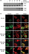
Examining the induction of Pa-T3S sensitivity in HL-60 cells. (a) Pa-T3S-resistant rHL-60 cells were treated with 20 nM TPA for the indicated time, prior to co-culture with Pa-ExoS-HA. Cells were lysed and fractionated, and the membrane fraction was resolved by SDS-PAGE and immunoblotted for ExoS and RalA to assay T3S effector translocation and function, and for PopB and PopD to assess T3S translocon membrane insertion. sn, P. aeruginosa culture supernatant used as molecular mass markers for ExoS, PopB and PopD. (b) The morphology of rHL-60 cells, T3S-sensitive sHL-60 cells and TPA-differentiated dHL-60 cells (differentiated for 12, 18 or 24 h) was examined by IF staining for tubulin (green) and actin (red). MT radiations developed during the early stages of Pa-T3S sensitivity and continued to develop in dHL-60 cells in association with actin-enriched projections and induction of Pa-T3S sensitivity. Images are representative of common phenotypes observed under the indicated experimental conditions in two or more experiments. Bar, 10 μm.
Fig. 4.
Fig. 4.
Examining adherence mechanisms and LE properties involved in host cell Pa-T3S sensitivity. (a) Diagram of major eukaryotic cell proteins mediating LE focal complex adherence. Proteins shown in solid lined boxes were found to mediate Pa-T3S sensitivity based on inhibition studies. To examine the role of focal complexes in Pa-T3S, HT-29 cells were seeded and allowed to adhere for 48 h (black bars), before treatment with Rac1 inhibitor (NSC23766) or Cdc42 inhibitor (secramine A), or were treated with inhibitors in the same manner immediately following seeding prior to establishment of adherence (grey bars). Cells were then co-cultured with Pa-ExoS-HA in the presence of inhibitors, and Pa-T3S sensitivity was quantified in cell lysates based on the efficiency of ExoS ADP ribosylation of RalA. The mean±sem of three independent experiments is presented. Significant decreases relative to non-drug-treated controls are indicated by *(P<0.05) or **(P<0.025). (b) To examine the role of LE properties in Pa-T3S sensitivity, HT-29 cells were treated with the indicated inhibitors, either 48 h after (black bars) or immediately following seeding (grey bars) and co-cultured with Pa-ExoS-HA and assayed for Pa-T3S sensitivity as in (a). Inhibitor treatments included: LatB, to disrupt actin; nocodazole, combined with incubation at 4 °C for 3 h, to destabilize/disrupt MTs; AS605240, to inhibit PI3Kγ; and LY294002, to inhibit all PI3K isoforms. Drug concentration relative to the efficiency of Pa-T3S effector translocation is shown as in (a), and the mean±sem of five to seven independent experiments are represented. Significant decreases in RalA ADP ribosylation relative to non-drug-treated controls are indicated by *(P<0.02) or **(P<0.002).
Fig. 5.
Fig. 5.
P. aeruginosa binding to LE of T3S-sensitive dHL-60 cells. HL-60 cells were differentiated with TPA for 18 h, then co-cultured with P. aeruginosa and stained for P. aeruginosa (blue) relative to LE-associated Rac or IQGAP1 (both green) and actin (red). Images show LE properties underlying P. aeruginosa binding and are representative of staining patterns obtained from two or more independent experiments. Bar, 10 μm.
Fig. 6.
Fig. 6.
P. aeruginosa binding to LE of HT-29 cells. P. aeruginosa binding to T3S-sensitive or T3S-resistant HT-29 cells was assessed following co-culture with Pa-ExoS-HA and staining for IQGAP1 (green), actin (red) and P. aeruginosa (blue). Left panel (lower magnification) shows the frequency of P. aeruginosa association with the LE of clusters of T3S-sensitive HT-29 cells. Bar, 20 μm. Middle panel (higher magnification) shows P. aeruginosa association with LE lamellipodia. Right image shows the more random and intercellular association of P. aeruginosa with HT-29 cells treated with 0.2 μg PFO ml−1 to cause loss of T3S sensitivity. Results are representative images obtained in two or three independent experiments. Bars, 10 μm.
Fig. 7.
Fig. 7.
Model for the role of eukaryotic cell properties in Pa-T3S. Our model predicts that alterations in membrane properties associated with F-actin polymerization at the LE induce forces on membranes that are required for PopB (B) and PopD (D) translocon insertion and function. V, PcrV, (PscF) T3S needle protein. The role of specific LE proteins in Pa-T3S is described in the text. Pa, P. aeruginosa.

Similar articles

Cited by

References

    1. Allmond, L. R., Karaca, T. J., Nguyen, V. N., Nguyen, T., Wiener-Kronish, J. P. & Sawa, T. (2003). Protein binding between PcrG-PcrV and PcrH-PopB/PopD encoded by the pcrGVH-popBD operon of the Pseudomonas aeruginosa type III secretion system. Infect Immun 71, 2230–2233. - PMC - PubMed
    1. Barber, D. F., Bartolomé, A., Hernandez, C., Flores, J. M., Redondo, C., Fernandez-Arias, C., Camps, M., Rückle, T., Schwarz, M. K. & other authors (2005). PI3Kγ inhibition blocks glomerulonephritis and extends lifespan in a mouse model of systemic lupus. Nat Med 11, 933–935. - PubMed
    1. Bashour, A. M., Fullerton, A. T., Hart, M. J. & Bloom, G. S. (1997). IQGAP1, a Rac- and Cdc42-binding protein, directly binds and cross-links microfilaments. J Cell Biol 137, 1555–1566. - PMC - PubMed
    1. Camps, M., Rückle, T., Ji, H., Ardissone, V., Rintelen, F., Shaw, J., Ferrandi, C., Chabert, C., Gillieron, C. & other authors (2005). Blockade of PI3K suppresses joint inflammation and damage in mouse models of rheumatoid arthritis. Nat Med 11, 936–943. - PubMed
    1. Collins, S. J., Ruscetti, F. W., Gallagher, R. E. & Gallo, R. C. (1978). Terminal differentiation of human promyelocytic leukemia cells induced by dimethyl sulfoxide and other polar compounds. Proc Natl Acad Sci U S A 75, 2458–2462. - PMC - PubMed

Publication types

MeSH terms