Comparative Study
. 2010 Feb;77(2):298-310.
doi: 10.1124/mol.109.061820. Epub 2009 Nov 12.

Cisplatin enhances protein kinase R-like endoplasmic reticulum kinase- and CD95-dependent melanoma differentiation-associated gene-7/interleukin-24-induced killing in ovarian carcinoma cells

Affiliations
Comparative Study

Cisplatin enhances protein kinase R-like endoplasmic reticulum kinase- and CD95-dependent melanoma differentiation-associated gene-7/interleukin-24-induced killing in ovarian carcinoma cells

Adly Yacoub et al. Mol Pharmacol. 2010 Feb.

Abstract

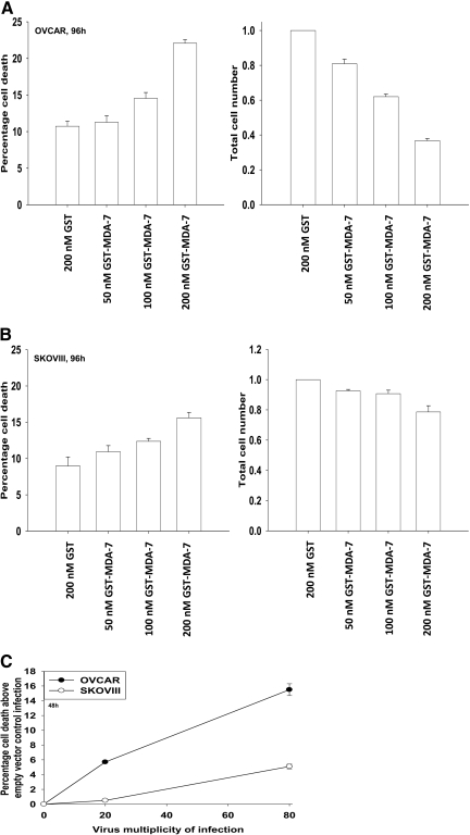
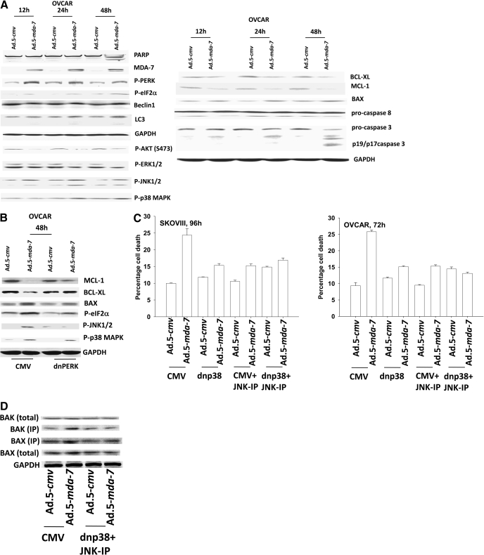
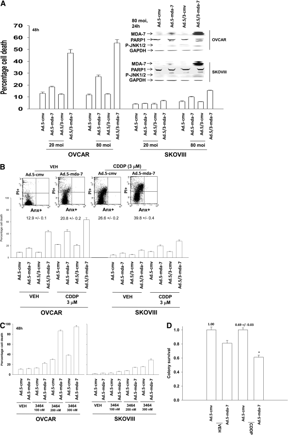
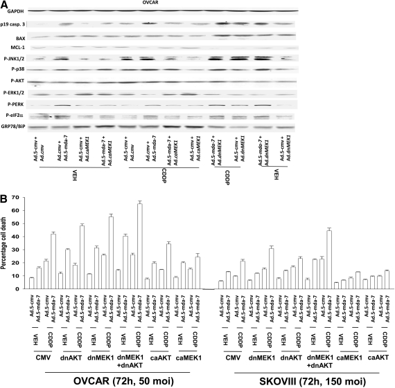
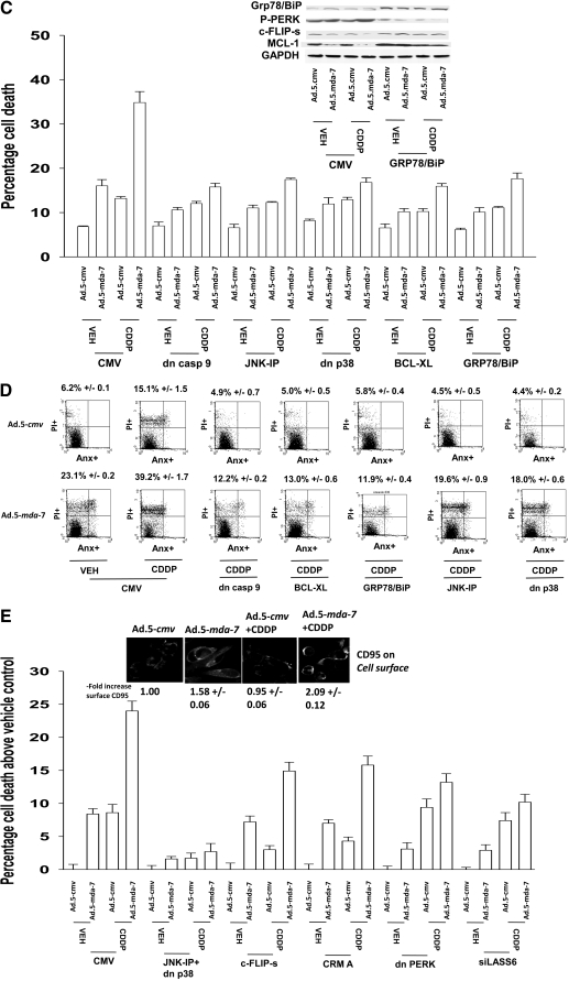
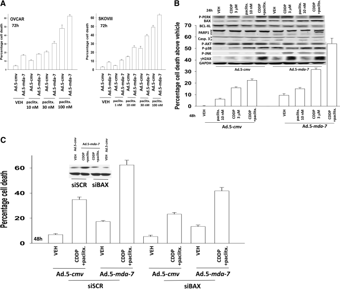
Melanoma differentiation associated gene-7/interleukin 24 (mda-7/IL-24) is a unique interleukin (IL)-10 family cytokine displaying selective apoptosis-inducing activity in transformed cells without harming normal cells. The present studies focused on defining the mechanism(s) by which recombinant adenoviral delivery of MDA-7/IL-24 inhibits cell survival of human ovarian carcinoma cells. Expression of MDA-7/IL-24 induced phosphorylation of protein kinase R-like endoplasmic reticulum kinase (PERK) and eukaryotic initiation factor2alpha (eIF2alpha). In a PERK-dependent fashion, MDA-7/IL-24 reduced ERK1/2 and AKT phosphorylation and activated c-Jun NH(2)-terminal kinase (JNK) 1/2 and p38 mitogen-activated protein kinase (MAPK). MDA-7/IL-24 reduced MCL-1 and BCL-XL and increased BAX levels via PERK signaling; cell-killing was mediated via the intrinsic pathway, and cell killing was primarily necrotic as judged using Annexin V/propidium iodide staining. Inhibition of p38 MAPK and JNK1/2 abolished MDA-7/IL-24 toxicity and blocked BAX and BAK activation, whereas activation of mitogen-activated extracellular-regulated kinase (MEK) 1/2 or AKT suppressed enhanced killing and JNK1/2 activation. MEK1/2 signaling increased expression of the MDA-7/IL-24 and PERK chaperone BiP/78-kDa glucose regulated protein (GRP78), and overexpression of BiP/GRP78 suppressed MDA-7/IL-24 toxicity. MDA-7/IL-24-induced LC3-green fluorescent protein vesicularization and processing of LC3; and knockdown of ATG5 suppressed MDA-7/IL-24-mediated toxicity. MDA-7/IL-24 and cisplatin interacted in a greater than additive fashion to kill tumor cells that was dependent on a further elevation of JNK1/2 activity and recruitment of the extrinsic CD95 pathway. MDA-7/IL-24 toxicity was enhanced in a weak additive fashion by paclitaxel; paclitaxel enhanced MDA-7/IL-24 + cisplatin lethality in a greater than additive fashion via BAX. Collectively, our data demonstrate that MDA-7/IL-24 induces an endoplasmic reticulum stress response that activates multiple proapoptotic pathways, culminating in decreased ovarian tumor cell survival.


Figures

Fig. 1.
Fig. 1.
MDA-7/IL-24 causes a dose-dependent induction of growth arrest and OCC death. A and B, OVCAR cells and SKOVIII cells were treated 24 h after plating with GST-MDA-7 (0–200 nM). At the indicated time point after GST-MDA-7 treatment (96 h), cell viability was determined by trypan blue exclusion assay, and cell numbers were determined using hemocytometer (± S.E.M., n = 3). C, OVCAR and SKOVIII cells were infected with type 5 recombinant adenoviruses empty vector (Ad.5-cmv) or to express MDA-7/IL-24 (Ad.5-mda-7) at an m.o.i. of 20 or 80. Cells were isolated 48 h after exposure, and the loss of cell viability above empty vector Ad.5-cmv infection was determined by using Annexin V propidium iodide staining assays in triplicate using a flow cytometer (± S.E.M., n = 3).
Fig. 2.
Fig. 2.
MDA-7/IL-24 promotes transformed cell killing through ER stress signaling and activation of the JNK/p38 MAPK pathways. A, OVCAR cells were infected with Ad.5-cmv or Ad.5-mda-7 at an m.o.i. of 50. Cells were isolated 12–48 h after infection and processed for SDS-PAGE and immunoblotting against the indicated proteins (n = 3). B, OVCAR cells were transfected with empty vector plasmid (CMV) or with a plasmid to express dominant-negative PERK. Twelve hours after transfection, cells were infected with Ad.5.cmv or Ad.5.mda-7 at an m.o.i. of 50. Cells were isolated 48 h after infection and processed for SDS-PAGE and immunoblotting against the indicated proteins (n = 3). C, OVCAR and SKOV3 cells were infected at an m.o.i. of 50 and 100, respectively, with Ad.5-cmv or Ad.5-mda-7. In parallel as indicated, cells were infected with a virus to express dominant-negative p38 MAPK. Twenty-four hours after infection, cells were treated with vehicle or with JNK-IP (10 μM). At the indicated time points in the graphs, cells were isolated, and the loss of cell viability was determined by trypan blue exclusion assays in triplicate (±S.E.M., n = 3). D, OVCAR cells were infected with viruses and treated with JNK-IP as in C. Forty-eight hours after infection, cells were isolated, and portions of the lysates were subjected to immunoprecipitation for isolation of activated forms of BAX and BAK. The precipitates were subjected to SDS-PAGE alongside an equal portion of the total cell lysates, and blotting was performed to determine the levels of BAX, BAK, and GAPDH (n = 2). E, OVCAR and SKOVIII cells were infected at an m.o.i. of 50 and 100, respectively, with Ad.5-cmv or Ad.5-mda-7. In parallel as indicated, cells were infected with recombinant adenoviruses to express activated and dominant-negative forms of AKT and MEK1 or with viruses to express apoptosis inhibitory proteins (c-FLIP-s, BCL-XL, dominant-negative caspase 9). Treatment with LY294002 (10 μM) occurred 24 h after virus infection. At the indicated time points in the graphs, cells were isolated, and the loss of cell viability was determined by trypan blue exclusion assays in triplicate (± S.E.M., n = 3). F, OVCAR cells were transfected as indicated with expression plasmids (empty vector CMV, dominant-negative PERK, BiP/GRP78, MCL-1) or with siRNA (siSCR, siATG5) and 12 h after transfection, cells were infected with Ad.5-cmv or Ad.5-mda-7 at an m.o.i. of 50. Cells were isolated 72 h after infection, and the loss of cell viability was determined by trypan blue exclusion assays in triplicate (± S.E.M., n = 3).
Fig. 2.
Fig. 2.
MDA-7/IL-24 promotes transformed cell killing through ER stress signaling and activation of the JNK/p38 MAPK pathways. A, OVCAR cells were infected with Ad.5-cmv or Ad.5-mda-7 at an m.o.i. of 50. Cells were isolated 12–48 h after infection and processed for SDS-PAGE and immunoblotting against the indicated proteins (n = 3). B, OVCAR cells were transfected with empty vector plasmid (CMV) or with a plasmid to express dominant-negative PERK. Twelve hours after transfection, cells were infected with Ad.5.cmv or Ad.5.mda-7 at an m.o.i. of 50. Cells were isolated 48 h after infection and processed for SDS-PAGE and immunoblotting against the indicated proteins (n = 3). C, OVCAR and SKOV3 cells were infected at an m.o.i. of 50 and 100, respectively, with Ad.5-cmv or Ad.5-mda-7. In parallel as indicated, cells were infected with a virus to express dominant-negative p38 MAPK. Twenty-four hours after infection, cells were treated with vehicle or with JNK-IP (10 μM). At the indicated time points in the graphs, cells were isolated, and the loss of cell viability was determined by trypan blue exclusion assays in triplicate (±S.E.M., n = 3). D, OVCAR cells were infected with viruses and treated with JNK-IP as in C. Forty-eight hours after infection, cells were isolated, and portions of the lysates were subjected to immunoprecipitation for isolation of activated forms of BAX and BAK. The precipitates were subjected to SDS-PAGE alongside an equal portion of the total cell lysates, and blotting was performed to determine the levels of BAX, BAK, and GAPDH (n = 2). E, OVCAR and SKOVIII cells were infected at an m.o.i. of 50 and 100, respectively, with Ad.5-cmv or Ad.5-mda-7. In parallel as indicated, cells were infected with recombinant adenoviruses to express activated and dominant-negative forms of AKT and MEK1 or with viruses to express apoptosis inhibitory proteins (c-FLIP-s, BCL-XL, dominant-negative caspase 9). Treatment with LY294002 (10 μM) occurred 24 h after virus infection. At the indicated time points in the graphs, cells were isolated, and the loss of cell viability was determined by trypan blue exclusion assays in triplicate (± S.E.M., n = 3). F, OVCAR cells were transfected as indicated with expression plasmids (empty vector CMV, dominant-negative PERK, BiP/GRP78, MCL-1) or with siRNA (siSCR, siATG5) and 12 h after transfection, cells were infected with Ad.5-cmv or Ad.5-mda-7 at an m.o.i. of 50. Cells were isolated 72 h after infection, and the loss of cell viability was determined by trypan blue exclusion assays in triplicate (± S.E.M., n = 3).
Fig. 3.
Fig. 3.
A tropism-modified type 5/type 3 virus infects ovarian cancer cells more readily than a type 5 virus and enhances cisplatin toxicity. A, OVCAR and SKOVIII cells were infected with Ad.5-cmv or Ad.5-mda-7 or with tropism-modified viruses Ad.5/3-cmv or Ad.5/3-mda-7 at an m.o.i. of 20 or 80, as indicated. Cells were isolated 48 h after infection, and the loss of cell viability was determined by trypan blue exclusion assays in triplicate (± S.E.M., n = 3). Inset, cells were infected with the indicated viruses, and 24 h after infection, cells were isolated to determine the expression of MDA-7/IL-24 and the cleavage status of PARP1. B, OVCAR and SKOVIII cells were infected with Ad.5-cmv or Ad.5-mda-7 or with tropism-modified viruses Ad.5/3-cmv or Ad.5/3-mda-7 at an m.o.i. of 80. Twenty-four h after infection, cells were treated with vehicle or cisplatin (CDDP, 3 μM). Seventy-two hours after infection, cells were isolated, and the loss of cell viability was determined by trypan blue exclusion assays in triplicate (± S.E.M., n = 3). Inset, identical portions of OVCAR cells, infected as indicated, were isolated 72 h after exposure, and the loss of cell viability was determined by using Annexin V/propidium iodide staining assays in triplicate using a flow cytometer (± S.E.M., n = 3). C, OVCAR and SKOVIII cells were infected with Ad.5-cmv or Ad.5-mda-7 at an m.o.i. of 80, and 24 h after infection, cells were treated with vehicle or BBR3464 (BBR3464, 100–300 nM). Forty-eight hours after infection, cells were isolated, and the loss of cell viability was determined by trypan blue exclusion assays in triplicate (± S.E.M., n = 3). D, OVCAR cells were plated in sextuplicate as single cells were infected with Ad.5-cmv or Ad.5-mda-7 at an m.o.i. of 80, and 24 h after infection, cells were treated with vehicle or cisplatin (CDDP, 3 μM). The media were changed to drug-free media 48 h after CDDP treatment, and colonies were permitted to form for 10 to 14 days (± S.E.M., n = 2).
Fig. 4.
Fig. 4.
Ad.5-mda-7 + cisplatin toxicity in OCCs is dependent on PERK, JNK and CD95 signaling. A, OVCAR cells were infected with Ad.5-cmv or Ad.5-mda-7 in parallel with Ad.cmv, Ad.MEK1 EE, or Ad.dnMEK1. Twenty-four hours after infection, cells were treated with vehicle or CDDP (3 μM). Forty-eight hours after infection, cells were isolated and processed for SDS-PAGE and immunoblotting against the indicated proteins (n = 3). B, OVCAR and SKOVIII cells were infected with the indicated viruses at the stated m.o.i., and 24 h after infection, cells were treated with vehicle or cisplatin (CDDP, 3 μM). Cells were isolated 72 h after infection, and the loss of cell viability was determined by trypan blue exclusion assays in triplicate (± S.E.M., n = 3). C, OVCAR cells were infected with Ad.5-cmv or Ad.5-mda-7 at an m.o.i. of 80 in parallel with viruses to express dominant-negative caspase 9, BCL-XL, or dominant-negative p38 MAPK. In parallel, cells were transfected with empty vector plasmid or plasmid to express Grp78/BiP. Twenty-four hours after infection as indicated, cells were treated with JNK-IP (10 μM) followed by cisplatin treatment (CDDP, 3 μM). Cells were isolated 72 h after infection, and the loss of cell viability was determined by trypan blue exclusion assays in triplicate (± S.E.M., n = 3). Inset, cells infected to express MDA-7/IL-24 and/or BiP/GRP78 were isolated 48 h after infection and processed for SDS-PAGE and blotting to determine the expression of MCL-1, c-FLIP-s, and GAPDH and the phosphorylation of PERK. D, OVCAR cells were infected and treated with cisplatin in a fashion identical with that described in C. Cells were isolated 72 h after infection, and the loss of cell viability was determined by using Annexin V/propidium iodide staining assays in triplicate using a flow cytometer (± S.E.M., n = 3). E, OVCAR cells were infected with caspase 8 inhibitors (c-FLIP-s, CRM A) or dominant-negative p38 MAPK or were transfected to express dominant-negative PERK or to knockdown ceramide synthase 6 (LASS6). Twenty-four hours after infection, cells where indicated were treated with JNK-IP (10 μM). Cells were isolated 72 h after infection, and the loss of cell viability was determined by trypan blue exclusion assays in triplicate (± S.E.M., n = 3). Inset, OVCAR cells growing in glass-chambered slides in triplicate were infected to express MDA-7/IL-24 and were treated 24 h after infection with CDDP (3 μM) for 6 h. Cells were fixed but not permeabilized, and the levels of cell surface CD95 was determined by immunohistochemistry (± S.E.M., n = 2). dn, dominant negative.
Fig. 4.
Fig. 4.
Ad.5-mda-7 + cisplatin toxicity in OCCs is dependent on PERK, JNK and CD95 signaling. A, OVCAR cells were infected with Ad.5-cmv or Ad.5-mda-7 in parallel with Ad.cmv, Ad.MEK1 EE, or Ad.dnMEK1. Twenty-four hours after infection, cells were treated with vehicle or CDDP (3 μM). Forty-eight hours after infection, cells were isolated and processed for SDS-PAGE and immunoblotting against the indicated proteins (n = 3). B, OVCAR and SKOVIII cells were infected with the indicated viruses at the stated m.o.i., and 24 h after infection, cells were treated with vehicle or cisplatin (CDDP, 3 μM). Cells were isolated 72 h after infection, and the loss of cell viability was determined by trypan blue exclusion assays in triplicate (± S.E.M., n = 3). C, OVCAR cells were infected with Ad.5-cmv or Ad.5-mda-7 at an m.o.i. of 80 in parallel with viruses to express dominant-negative caspase 9, BCL-XL, or dominant-negative p38 MAPK. In parallel, cells were transfected with empty vector plasmid or plasmid to express Grp78/BiP. Twenty-four hours after infection as indicated, cells were treated with JNK-IP (10 μM) followed by cisplatin treatment (CDDP, 3 μM). Cells were isolated 72 h after infection, and the loss of cell viability was determined by trypan blue exclusion assays in triplicate (± S.E.M., n = 3). Inset, cells infected to express MDA-7/IL-24 and/or BiP/GRP78 were isolated 48 h after infection and processed for SDS-PAGE and blotting to determine the expression of MCL-1, c-FLIP-s, and GAPDH and the phosphorylation of PERK. D, OVCAR cells were infected and treated with cisplatin in a fashion identical with that described in C. Cells were isolated 72 h after infection, and the loss of cell viability was determined by using Annexin V/propidium iodide staining assays in triplicate using a flow cytometer (± S.E.M., n = 3). E, OVCAR cells were infected with caspase 8 inhibitors (c-FLIP-s, CRM A) or dominant-negative p38 MAPK or were transfected to express dominant-negative PERK or to knockdown ceramide synthase 6 (LASS6). Twenty-four hours after infection, cells where indicated were treated with JNK-IP (10 μM). Cells were isolated 72 h after infection, and the loss of cell viability was determined by trypan blue exclusion assays in triplicate (± S.E.M., n = 3). Inset, OVCAR cells growing in glass-chambered slides in triplicate were infected to express MDA-7/IL-24 and were treated 24 h after infection with CDDP (3 μM) for 6 h. Cells were fixed but not permeabilized, and the levels of cell surface CD95 was determined by immunohistochemistry (± S.E.M., n = 2). dn, dominant negative.
Fig. 5.
Fig. 5.
Paclitaxel enhances the toxicity of Ad.5-mda-7 + cisplatin in a greater than additive fashion. A, OVCAR and SKOVIII cells were infected with Ad.5-cmv or Ad.5-mda-7 at an m.o.i. of 80. Twenty-four hours after infection, as indicated, cells were treated with paclitaxel (paclitx., 0–100 nM). Cells were isolated 48 h after infection, and the loss of cell viability was determined by trypan blue exclusion assays in triplicate (± S.E.M., n = 3). B, OVCAR cells were infected with Ad.5-cmv or Ad.5-mda-7 at an m.o.i. of 80. Twenty-four hours after infection, as indicated, cells were treated with paclitaxel (paclitx., 10 nM) and/or cisplatin (CDDP, 3 μM). Cells were isolated 48 h after infection, and the loss of cell viability was determined by trypan blue exclusion assays in triplicate (± S.E.M., n = 3). Top inset, cells were isolated 24 h after virus infection and processed for SDS-PAGE and immunoblotting against the indicated proteins to determine expression/phosphorylation (n = 2). C, OVCAR cells were transfected with scrambled siRNA (siSCR) or an siRNA to knock down BAX expression and 24 h later were infected with Ad.5-cmv or Ad.5-mda-7 at an m.o.i. of 80. Twenty-four hours after infection, as indicated, cells were treated with paclitaxel (paclitx., 10 nM) and cisplatin (CDDP, 3 μM). Cells were isolated 48 h after infection, and the loss of cell viability was determined by trypan blue exclusion assays in triplicate (± S.E.M., n = 3).

Similar articles

Cited by

References

    1. Caudell EG, Mumm JB, Poindexter N, Ekmekcioglu S, Mhashilkar AM, Yang XH, Retter MW, Hill P, Chada S, Grimm EA. (2002) The protein product of the tumor suppressor gene, melanoma differentiation-associated gene 7, exhibits immunostimulatory activity and is designated IL-24. J Immunol 168: 6041–6046 - PubMed
    1. Chada S, Bocangel D, Ramesh R, Grimm EA, Mumm JB, Mhashilkar AM, Zheng M. (2005) mda-7/IL24 kills pancreatic cancer cells by inhibition of the Wnt/PI3K signaling pathways: identification of IL-20 receptor-mediated bystander activity against pancreatic cancer. Mol Ther 11: 724–733 - PubMed
    1. Coleman RL, Sood AK. (2006) Historical progress in the initial management of ovarian cancer: intraperitoneal chemotherapy. Curr Oncol Rep 8: 455–464 - PubMed
    1. Cunningham CC, Chada S, Merritt JA, Tong A, Senzer N, Zhang Y, Mhashilkar A, Parker K, Vukelja S, Richards D, et al. (2005) Clinical and local biological effects of an intratumoral injection of mda-7 (IL24; INGN 241) in patients with advanced carcinoma: a phase I study. Mol Ther 11: 149–159 - PubMed
    1. Dash R, Dmitriev IP, Su ZZ, Bhutia SJ, Azab B, Vozhilla N, Yacoub A, Dent P, Curiel DT, Sarkar D, et al. (2010) Enhanced delivery of mda-7/IL-24 using a serotype chimeric adenovirus (Ad.5/3) improves therapeutic efficacy in low CAR prostate cancer cells. Cancer Gene Ther, in press - PubMed

Publication types

MeSH terms