. 2010 Jan 22;285(4):2758-70.
doi: 10.1074/jbc.M109.066845. Epub 2009 Nov 12.

Jmjd1a demethylase-regulated histone modification is essential for cAMP-response element modulator-regulated gene expression and spermatogenesis

Affiliations

Jmjd1a demethylase-regulated histone modification is essential for cAMP-response element modulator-regulated gene expression and spermatogenesis

Zhaoliang Liu et al. J Biol Chem. .

Abstract

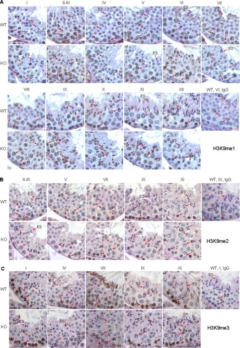
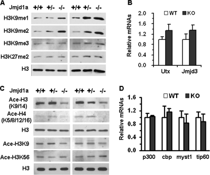
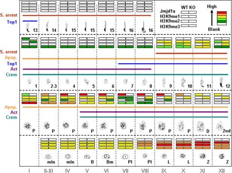
Spermatogenesis, a fundamental process in the male reproductive system, requires a series of tightly controlled epigenetic and genetic events in germ cells ranging from spermatogonia to spermatozoa. Jmjd1a is a key epigenetic regulator expressed in the testis. It specifically demethylates mono- and di-methylated histone H3 lysine 9 (H3K9me1 and H3K9me2) but not tri-methylated H3K9 (H3K9me3). In this study, we generated a Jmjd1a antibody for immunohistochemistry and found Jmjd1a was specifically produced in pachytene and secondary spermatocytes. Disruption of the Jmjd1a gene in mice significantly increased H3K9me1 and H3K9me2 levels in pachytene spermatocytes and early elongating spermatids without affecting H3K9me3 levels. Concurrently, the levels of histone acetylation were decreased in Jmjd1a knock-out germ cells. This suggests Jmjd1a promotes transcriptional activation by lowering histone methylation and increasing histone acetylation. Interestingly, the altered histone modifications in Jmjd1a-deficient germ cells caused diminished cAMP-response element modulator (Crem) recruitment to chromatin and decreased expression of the Crem coactivator Act and their target genes Tnp1 (transition protein 1), Tnp2, Prm1 (protamine 1), and Prm2, all of which are essential for chromatin condensation in spermatids. In agreement with these findings, Jmjd1a deficiency caused extensive germ cell apoptosis and blocked spermatid elongation, resulting in severe oligozoospermia, small testes, and infertility in male mice. These results indicate that the Jmjd1a-controlled epigenetic histone modifications are crucial for Crem-regulated gene expression and spermatogenesis.


Figures

FIGURE 1.
FIGURE 1.
Generation of Jmjd1a−/− mice. A, gene-targeting strategy used to generate Jmjd1a knock-out allele in ES cells. The relationships among the 3′ region of the Jmjd1a gene, the targeting vector, and the targeted Jmjd1a allele are sketched. The two black bars under the Jmjd1a locus indicate the 5′ and 3′ DNA probes for Southern blot. The gray bars under the Jmjd1a locus and the targeted allele indicate the DNA fragments amplified by PCR for genotyping. B, identification of targeted ES clones by Southern blot. ES cell DNA was digested by XhoI and EcoRV for Southern blot using 5′ probe and by EcoRV for Southern blot using 3′ probe as indicated in A. The 5′ probe detected a 10.9-kb fragment from the WT (+) Jmjd1a allele and a 7.3-kb fragment from the targeted (−) Jmjd1a allele. The 3′ probe detected a 11.4-kb fragment from the WT Jmjd1a allele and a 8.1-kb fragment from the targeted Jmjd1a allele. C, genotype analysis of mice by PCR. PCRs were performed using mouse genomic DNA and allele-specific primer pairs. The upper band (226 bp) and the lower band (120 bp) represent WT and targeted Jmjd1a alleles, respectively. D, Western blot. Testis lysates were prepared from Jmjd1a heterozygous (+/−) and knock-out (−/−) mice. Western blot was performed using Jmjd1a antibody. β-Actin served as a loading control.
FIGURE 2.
FIGURE 2.
Testis weight, sperm count, and Jmjd1a distribution in the testis. A, photographs of testes and the ratios of testis weight (T.W.) to body weight (B.W.) obtained from 12-week-old WT and Jmjd1a−/− (KO) mice. S.D., standard deviation; N., number; p value (<0.01), calculated by unpaired t test. B, hematoxylin and eosin-stained sections of the caudal epididymides prepared from 12-week-old WT and Jmjd1a−/− (KO) mice and the average sperm number collected from each epididymis in WT and KO mice. The statistical analysis was performed with unpaired t test. S, spermatozoa; L, lumen. C, Jmjd1a IHC. Paraffin sections were prepared from the testes of 7-week-old mice and used for IHC. Brown color indicates Jmjd1a immunoreactivity. The sections were counterstained with periodic acid-Schiff and hematoxylin. The section from a Jmjd1a−/− (KO) mouse served as a negative control. The seminiferous stages were labeled from I to XII. Sg, spermatogonia; Pl, preleptotene; L, leptotene; Z, zygotene; P, pachytene; 2nd, secondary spermatocyte; RS, round spermatid; ES, elongating spermatid; SC, Sertoli cell; LC, Leydig cell. D, immunofluorescence staining of chromosomes in a pachytene spermatocyte with Jmjd1a antibodies and propidium iodide (PI).
FIGURE 3.
FIGURE 3.
Stage-specific H3K9 methylation in the testes of WT mice and increased H3K9me1 and H3K9me2 in the testes of Jmjd1a−/− (KO) mice. Testis sections were prepared from 7-week-old WT and Jmjd1a−/− (KO) mice. The stages of seminiferous epithelium are indicated. Refer to the legend for Fig. 2 for abbreviations of cell type nomenclatures. Substitution of nonimmune IgG for specific antibodies was performed as negative controls. A, H3K9me1 IHC (brown color) in the testes of WT and KO mice. B, H3K9me2 IHC (brown color) in the testes of WT and KO mice. C, H3K9me3 IHC (brown color) in the testes of WT and KO mice.
FIGURE 4.
FIGURE 4.
Jmjd1a deficiency increases histone methylation and decreases histone acetylation in the testis. A, Western blot analyses of H3K9me1, H3K9me2, H3K9me3, H3K27me2, and total H3 in the testes of 12-week-old WT (+/+), heterozygous (+/−), and KO (−/−) Jmjd1a mice. Two independent sets of samples were assayed. Total H3 levels served as loading controls. B, Utx and Jmjd3 mRNA levels measured by real time RT-PCR. Total RNA samples were prepared from the testes of 12-week-old WT (n = 3) and KO (n = 3) mice. Each sample was assayed twice. The level of mRNA was normalized to 18 S level of the same sample. Data have no statistical difference and are presented as mean ± S.D. C, Western blot analyses of acetylated H3 (Ace-H3K9/14), H4 (Ace-H4K5/8/12/16), H3K9, and H3K56. Total H3 served as loading control. Two independent sets of samples were assayed. The first three testis samples used in A and C were the same, so the total H3 was the same. D, real time RT-PCR measurements of p300, Cbp, Myst1, and Tip60 mRNA levels. RNA samples and analytical methods were described in B.
FIGURE 5.
FIGURE 5.
Jmjd1a deficiency decreases Act expression and diminishes Crem-mediated gene expression. A, relative Crem and Act mRNA expression levels in the testes of 12-week-old WT and Jmjd1a−/− (KO) mice (n = 3 for each group). **, p < 0.01 by unpaired t test. B, cAMP-response elements (CRE) for Crem binding in the proximal regions of the Tnp1 and Odf1 promoters are indicated. Exon 1 and exon 2 (E2) also are indicated. The DNA fragments (a–c) that were amplified by PCR in ChIP assays are marked. C, ChIP assays for regions a and b of the Tnp1 promoter and c of the Odf1 promoter (B). Germ cells were isolated from the testes of WT and Jmjd1a−/− (KO) mice. Input represents 20% of materials used for Crem ChIP. ChIP assays were performed using antibodies against H3K9me1, H3K9me2, H3K9me3, and Crem as indicated. Substitution of nonimmune IgG for specific antibodies served as negative controls. D, expression levels of Crem and Act target genes in the testes of 12-week-old WT and Jmjd1a−/− (KO) mice (n = 3 for each group). Data are presented as mean ± S.D. *, p < 0.05 by unpaired t test. E, Western blot analysis of Tnp1 in the testes of 12-week-old WT (+/+), Jmjd1a+/−, and Jmjd1a−/− mice. β-Actin served as a loading control.
FIGURE 6.
FIGURE 6.
Ablation of Jmjd1a arrests spermiogenesis and causes apoptosis of developing germ cells. A, Jmjd1a deficiency blocked spermiogenesis at spermatid-elongating stages. Testis sections were prepared from 12-week-old WT and Jmjd1a−/− (KO) mice and stained with periodic acid-Schiff and hematoxylin. The stages of seminiferous epithelium are indicated from I to XII. Note the abnormal head morphologies of elongating spermatids at stages X–XII and stages I–IV and the drastically reduced number of elongated spermatids at stages X–VII in KO testes (green boxes). Sg, spermatogonia; Pl, preleptotene; L, leptotene; Z, zygotene; P, pachytene; 2nd, secondary spermatocyte; RS, round spermatid; ES, elongating spermatid; SC, Sertoli cell; LC, Leydig cell. B, toluidine blue-stained semi-thin sections of WT and Jmjd1a−/− (KO) mouse testes. Developmental steps of spermatids are indicated. acr, acrosome; DS, dorsal side; VS, ventral side. C, detection of apoptotic cells in the testis sections of 12-week-old WT and KO mice by terminal deoxynucleotidyltransferase nick end-labeling assay. Apoptotic cells exhibit brown nuclei in this assay. The lower panels are the enlarged pictures of the boxed regions in the upper panels. The asterisk indicates the small apoptotic nuclei of the elongating spermatids. D, quantitative analysis of apoptotic germ cells in the seminiferous epithelium of WT and KO testes. Three 12-week-old mice per genotype group and two sections per testis were analyzed. Apoptotic cells in group a (pre-pachytene germ cells), group b (pachytene, diplotene and secondary spermatocytes), and group c (spermatids) were counted and recorded separately. Data are presented as the average number (No.) of apoptotic cells per cross-sectioned seminiferous tubule. The Total bar graphs represent the sum of groups a–c. *, p < 0.05 by unpaired t test.
FIGURE 7.
FIGURE 7.
Temporal and spatial distributions of Jmjd1a, H3K9me1, H3K9me2, H3K9me3, Crem, Act, and Tnp1 in WT and Jmjd1a−/− mice during spermatogenesis and their relationships relevant to germ cell apoptosis (Apop.) and spermatid-elongating arrest (S. arrest) in Jmjd1a−/− mice. The 12 stages of seminiferous epithelium and the developmental steps of spermatogenesis are listed. The levels of Jmjd1a, H3K9me1, H3K9me2, and H3K9me3 in the testes of WT and Jmjd1a−/− (KO) mice are presented in color codes as indicated (upper right corner). The steps with Crem and Act proteins and Tnp1 mRNA expression are indicated. The steps with germ cell apoptosis and developmental arrest of elongating spermatids in Jmjd1a−/− mice are also indicated. mIn, mitotic intermediate spermatogonia; B, type B spermatogonia; Bm, mitotic type B spermatogonia; Pl, preleptotene cells; L, leptotene cells; Z, zygotene cells; P, pachytene cells; 2nd, secondary spermatocytes; 1–16, steps 1–16 spermatids.
FIGURE 8.
FIGURE 8.
A working model of Jmjd1a function in spermatogenesis.

Similar articles

Cited by

References

    1. Russell L. D., Ettlin R. A., Sinha-Hikim A. P., Clegg E. D. (1990) Histological and Histopathological Evaluation of the Testis, pp. 1–161, Cache River Press, Clearwater, FL
    1. Mali P., Kaipia A., Kangasniemi M., Toppari J., Sandberg M., Hecht N. B., Parvinen M. (1989) Reprod. Fertil. Dev. 1, 369–382 - PubMed
    1. Nantel F., Monaco L., Foulkes N. S., Masquilier D., LeMeur M., Henriksén K., Dierich A., Parvinen M., Sassone-Corsi P. (1996) Nature 380, 159–162 - PubMed
    1. Cho C., Willis W. D., Goulding E. H., Jung-Ha H., Choi Y. C., Hecht N. B., Eddy E. M. (2001) Nat. Genet. 28, 82–86 - PubMed
    1. Yu Y. E., Zhang Y., Unni E., Shirley C. R., Deng J. M., Russell L. D., Weil M. M., Behringer R. R., Meistrich M. L. (2000) Proc. Natl. Acad. Sci. U.S.A. 97, 4683–4688 - PMC - PubMed

Publication types

MeSH terms