. 2010 Jan 8;285(2):845-54.
doi: 10.1074/jbc.M109.045740. Epub 2009 Nov 12.

Ethanol enhances hepatitis C virus replication through lipid metabolism and elevated NADH/NAD+

Affiliations

Ethanol enhances hepatitis C virus replication through lipid metabolism and elevated NADH/NAD+

Scott Seronello et al. J Biol Chem. .

Abstract

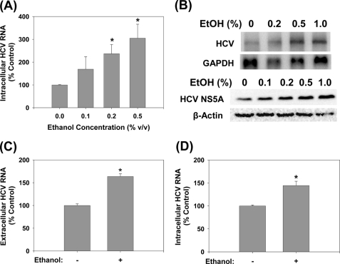
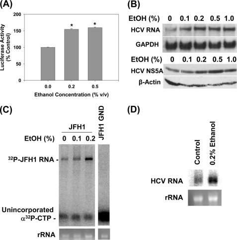
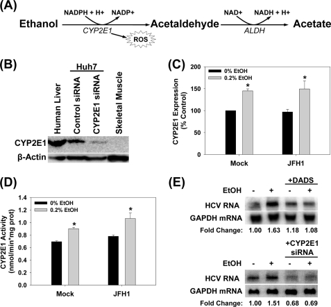
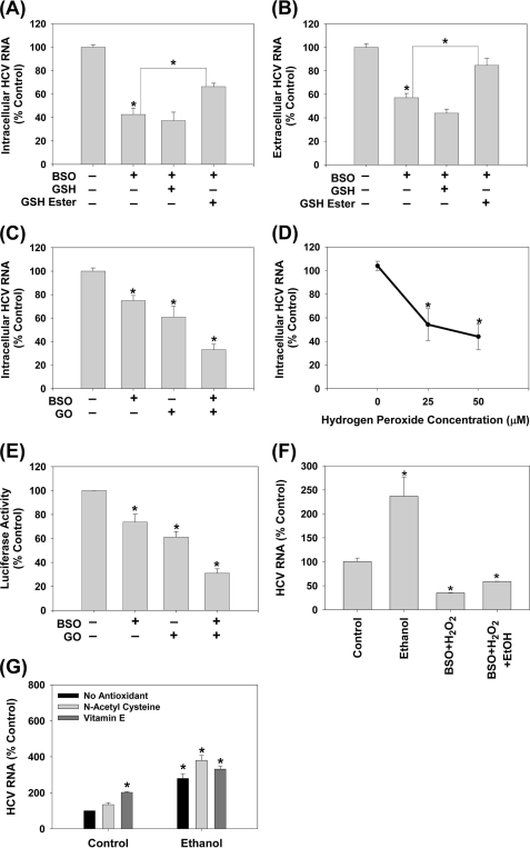
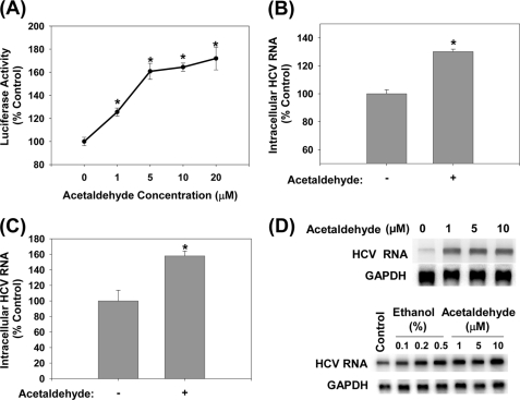
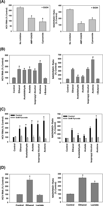
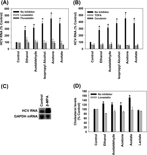
Ethanol has been suggested to elevate HCV titer in patients and to increase HCV RNA in replicon cells, suggesting that HCV replication is increased in the presence and absence of the complete viral replication cycle, but the mechanisms remain unclear. In this study, we use Huh7 human hepatoma cells that naturally express comparable levels of CYP2E1 as human liver to demonstrate that ethanol, at subtoxic and physiologically relevant concentrations, enhances complete HCV replication. The viral RNA genome replication is affected for both genotypes 2a and 1b. Acetaldehyde, a major product of ethanol metabolism, likewise enhances HCV replication at physiological concentrations. The potentiation of HCV replication by ethanol is suppressed by inhibiting CYP2E1 or aldehyde dehydrogenase and requires an elevated NADH/NAD(+) ratio. In addition, acetate, isopropyl alcohol, and concentrations of acetone that occur in diabetics enhance HCV replication with corresponding increases in the NADH/NAD(+). Furthermore, inhibiting the host mevalonate pathway with lovastatin or fluvastatin and fatty acid synthesis with 5-(tetradecyloxy)-2-furoic acid or cerulenin significantly attenuates the enhancement of HCV replication by ethanol, acetaldehyde, acetone, as well as acetate, whereas inhibiting beta-oxidation with beta-mercaptopropionic acid increases HCV replication. Ethanol, acetaldehyde, acetone, and acetate increase the total intracellular cholesterol content, which is attenuated with lovastatin. In contrast, both endogenous and exogenous ROS suppress the replication of HCV genotype 2a, as previously shown with genotype 1b.

Conclusion: Therefore, lipid metabolism and alteration of cellular NADH/NAD(+) ratio are likely to play a critical role in the potentiation of HCV replication by ethanol rather than oxidative stress.


Figures

FIGURE 1.
FIGURE 1.
Ethanol increases JFH1 replication. Huh7 cells transfected with JFH1 RNA were analyzed for intracellular HCV RNA by (A) qRT-PCR (n = 6) or (B) Northern blots (n = 4) and for HCV NS5A protein content by Western blot (n = 3) (B, bottom panel) after 48 h of ethanol treatments. C, extracellular HCV RNA levels were analyzed by qRT-PCR for 0.2% ethanol treatments (n = 4). D, naïve Huh7 cells were inoculated with virus-containing medium and analyzed for HCV RNA after 48 h of 0.2% ethanol treatment (n = 4). *, indicates statistically significant difference for indicated sample sizes (p < 0.05).
FIGURE 2.
FIGURE 2.
Ethanol increases the replication of subgenomic JFH1 and Con1 replicon RNAs. A, Huh7 cells transfected with SgJFH1-Luc RNA were analyzed for luciferase activity after 48-h ethanol treatments (n = 3). B, stable Huh7 clones expressing SgCon1-Neo (SgPC2) were incubated with ethanol for 24 h and analyzed for HCV RNA, GAPDH mRNA, and NS5A and β-actin proteins (n = 3) by Northern and Western blots, respectively (n = 3). C and D, cytosolic lysates were prepared from (C) JFH1 and JFH1-GND RNA-transfected cells and (D) SgPC2 cells, after 5 h of ethanol treatment, these lysates were used to carry out in vitro replication assays (n = 3). Bottom panels show ethidium bromide staining of rRNA as the loading control. *, indicates statistically significant difference for indicated sample sizes (p < 0.05).
FIGURE 3.
FIGURE 3.
CYP2E1 expression in Huh7 cells. A, CYP2E1-dependent ethanol metabolism. B, human liver tissue, Huh7 cells transfected with 50 μm non-targeting control or CYP2E1 siRNA, and skeletal muscle tissue were analyzed for CYP2E1 protein content by Western blot (n = 3). C and D, mock- or JFH1-transfected Huh7 cells were incubated with or without 0.2% (v/v) ethanol for 48 h and analyzed for (C) CYP2E1 expression by Western blot (n = 3) and (D) CYP2E1-dependent p-nitrophenol hydroxylation activity (n = 3). E, SgPC2 cells were exposed to 0.2% ethanol ± 25 μm DADS for 24 h or transfected with 50 nm control or CYP2E1 siRNA for 24 h and then incubated with ethanol for 24 h and analyzed for HCV RNA by Northern blot (n = 3). *, indicates statistically significant difference for indicated sample sizes (p < 0.05).
FIGURE 4.
FIGURE 4.
Endogenous and exogenous ROS suppress HCV replication. JFH1-transfected Huh7 cells were treated with BSO with and without 2 mm GSH or GSH ester (A and B) (n = 3), GO + glucose with and without 16 h of pretreatment with 20 μm BSO (C) (n = 4), or bolus H2O2 (D) (n = 4) for 24 h. Then, JFH1 intracellular (A, C, D) and extracellular (B) HCV RNA levels were analyzed by qRT-PCR. E, Huh7 cells transfected with SgJFH1-Luc RNA were assayed for luciferase activity after 24 h treatment with 0.25 milliunits/ml glucose oxidase + glucose with and without the BSO pretreatment (n = 3). F, SgPC2 cells were treated with 0.2% ethanol ± H2O2 plus BSO for 24 h, and analyzed for HCV RNA and GAPDH mRNA by Northern blot. G, SgPC2 cells were treated for 24 h with ethanol ± 5 mm NAC or 0.5 μm Trolox (water-soluble vitamin E). Then, HCV RNA and GAPDH mRNA levels were monitored by Northern blot and quantified by densitometry (n = 3). *, indicates statistically significant difference for indicated sample sizes (p < 0.05).
FIGURE 5.
FIGURE 5.
Acetaldehyde increases intracellular HCV RNA. SgJFH1-Luc (A) and JFH1 RNA-transfected cells (B), Huh7.5 cells inoculated with JFH1 virions (C), SgPC2 and Clone B cells (D) were incubated with acetaldehyde for 24 h and analyzed for HCV RNA by Northern blot or qRT-PCR (n = 3). *, indicates statistically significant difference for indicated sample size (p < 0.05).
FIGURE 6.
FIGURE 6.
Role of NADH/NAD+ in the potentiation of HCV replication by ethanol, acetaldehyde, acetate, isopropyl alcohol, and acetone. SgPC2 cells, supporting Con1 subgenomic HCV RNA replication, were treated with (A) 0.2% ethanol ± 0.1 mm 4MP plus 25 μm DADS or 0.1 mm cyanamide (n = 3); (B) 0.2% ethanol, 5 μm acetaldehyde, 5 μm acetate, 0.2% isopropyl alcohol, 2 mm acetone, or 25 mm tert-butanol (n = 4); (C) 0.2% ethanol, 5 μm acetaldehyde, 5 μm acetate, 0.2% isopropyl alcohol, and 2 mm acetone, with and without 5 mm pyruvate (n = 3); or (D) 0.2% ethanol or 5 mm lactate for 3 h for NADH/NAD+ ratio measurement or 24 h for HCV RNA levels. HCV RNA levels were monitored by Northern blot (A–D, left panels). NADH/NAD+ ratios were measured by an enzymatic NADH recycling assay, as described under “Experimental Procedures” (A–D, right panels). Northern blots were quantified by densitometry. *, indicates statistically significant difference for indicated sample sizes (p < 0.05).
FIGURE 7.
FIGURE 7.
Role of lipogenesis in the enhancement of HCV replication by ethanol, acetaldehyde, isopropyl alcohol, acetone, and acetate. SgPC2 cells were treated for 24 h with (A and B) 0.2% ethanol, 5 μm acetaldehyde, 0.2% isopropyl alcohol, 2 mm acetone, 5 μm acetate ± 30 min pretreatment with (A) 5 μm lovastatin, 5 μm fluvastatin, (B) 5 μg/ml TOFA, 5 μg/ml cerulenin, or with (C) 2 mm β-mercaptopropionic acid (β-MPA). Then, HCV RNA levels were monitored by Northern blot and quantified by densitometry (n = 3). D, SgPC2 cells, treated for 24 h with ethanol, acetaldehyde, acetone, and acetate ± lovastatin, were monitored for cholesterol levels (n = 3). Lovastatin was activated, as described, before use (29). *, indicates statistically significant difference for indicated sample sizes (p < 0.05).

Similar articles

Cited by

References

    1. Jamal M. M., Morgan T. R. (2003) Best Pract. Res. Clin. Gastroenterol. 17, 649–662 - PubMed
    1. Sata M., Fukuizumi K., Uchimura Y., Nakano H., Ishii K., Kumashiro R., Mizokami M., Lau J. Y., Tanikawa K. (1996) J. Viral. Hepat. 3, 143–148 - PubMed
    1. Zhang T., Li Y., Lai J. P., Douglas S. D., Metzger D. S., O'Brien C. P., Ho W. Z. (2003) Hepatology 38, 57–65 - PubMed
    1. Seronello S., Sheikh M. Y., Choi J. (2007) Free Radic. Biol. Med. 43, 869–882 - PubMed
    1. Cromie S. L., Jenkins P. J., Bowden D. S., Dudley F. J. (1996) J. Hepatol. 25, 821–826 - PubMed

Publication types

MeSH terms