. 2009 Dec;175(6):2392-405.
doi: 10.2353/ajpath.2009.090114. Epub 2009 Nov 12.

Farnesoid X receptor critically determines the fibrotic response in mice but is expressed to a low extent in human hepatic stellate cells and periductal myofibroblasts

Affiliations

Farnesoid X receptor critically determines the fibrotic response in mice but is expressed to a low extent in human hepatic stellate cells and periductal myofibroblasts

Peter Fickert et al. Am J Pathol. 2009 Dec.

Erratum in

  • Am J Pathol. 2010 May;176(5):2581

Abstract

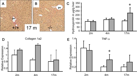
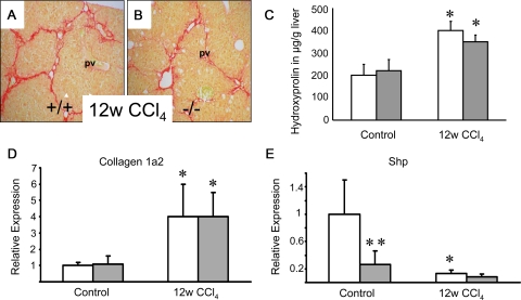
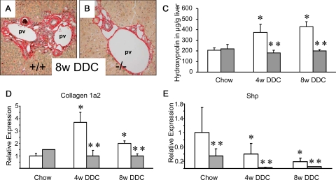
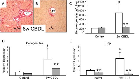
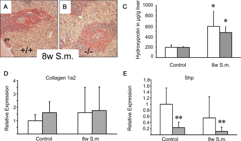
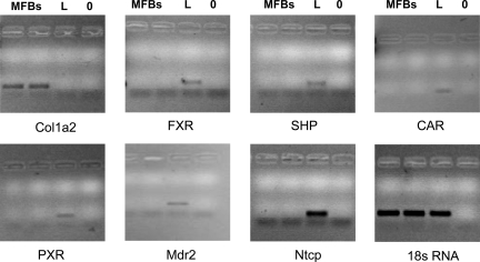
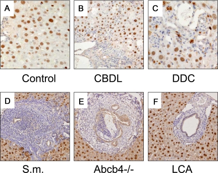
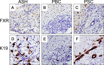
The nuclear bile acid receptor, farnesoid X receptor (FXR), may play a pivotal role in liver fibrosis. We tested the impact of genetic FXR ablation in four different mouse models. Hepatic fibrosis was induced in wild-type and FXR knock-out mice (FXR(-/-)) by CCl(4) intoxication, 3,5-diethoxycarbonyl-1,4-dihydrocollidine feeding, common bile duct ligation, or Schistosoma mansoni (S.m.)-infection. In addition, we determined nuclear receptor expression levels (FXR, pregnane X receptor (PXR), vitamin D receptor, constitutive androstane receptor (CAR), small heterodimer partner (SHP)) in mouse hepatic stellate cells (HSCs), portal myofibroblasts (MFBs), and human HSCs. Cell type-specific FXR protein expression was determined by immunohistochemistry in five mouse models and prototypic human fibrotic liver diseases. Expression of nuclear receptors was much lower in mouse and human HSCs/MFBs compared with total liver expression with the exception of vitamin D receptor. FXR protein was undetectable in mouse and human HSCs and MFBs. FXR loss had no effect in CCl(4)-intoxicated and S.m.-infected mice, but significantly decreased liver fibrosis of the biliary type (common bile duct ligation, 3,5-diethoxycarbonyl-1,4-dihydrocollidine). These data suggest that FXR loss significantly reduces fibrosis of the biliary type, but has no impact on non-cholestatic liver fibrosis. Since there is no FXR expression in HSCs and MFBs in liver fibrosis, our data indicate that these cells may not represent direct therapeutic targets for FXR ligands.


Figures

Figure 1
Figure 1
Longitudinal comparison between FXR−/−mice and wild-type controls. Sirius-red stain for collagen staining in naïve 17-month-old wild-type (+/+) (A) and FXR knock-out (−/−) mice (B). C: Quantification of liver fibrosis by determination of hepatic hydroxyproline content shows equal levels in both genotypes at 2 and 4 months of age and significantly increased levels in 17-month-old FXR−/− mice (gray bars). D: Equal collagen 1a2 mRNA expression at 2 and 17 months of age, and elevated levels in 4-month-old FXR−/−mice. E: Equal hepatic TNF-α mRNA expression levels in both genotypes at 2 and 17 months of age and significantly elevated levels in 4-month-old FXR−/−mice. *P < 0.05 (wild-type versus FXR−/− mice). Open bars, wild-type mice; gray bars, FXR knock-out mice. pv, portal vein. Original magnification for A, B, ×10.
Figure 2
Figure 2
Loss of FXR has no effect on liver fibrosis in CCl4-intoxicated mice. Sirius-red staining for collagen in 12 weeks CCl4-intoxicated wild-type (+/+) (A) and FXR knock-out (−/−) (B). A similar degree of Sirius red staining was observed in both wild-type (A) and FXR−/− (B) after CCl4-intoxicated. C: Quantification of liver fibrosis by determination of hepatic hydroxyproline content shows similar levels between genotypes. D: Significantly increased collagen 1a2 mRNA expression in 12w CCl4-intoxicated wild-type mice and FXR−/−mice. E: Significantly lower Shp mRNA expression levels in naïve FXR−/−mice and CCl4-intoxicated wild-type mice. Open bars, wild-type mice; gray bars, FXR knock-out mice. *P < 0.05 (naïve versus CCl4-intoxicated wild-type/FXR−/−), **P < 0.05 (WT vs. FXR−/−). pv, portal vein. Original magnification for A, B, ×10.
Figure 3
Figure 3
Loss of FXR significantly reduces liver fibrosis in 3,5-diethoxycarbonyl-1,4-dihydrocollidine (DDC)-fed mice. Sirius-red stain for collagen staining in mice fed 0.1% (w/w) DDC-supplemented diet for 8 weeks (A,B). Significantly increased Sirius-red staining in DDC-fed wild-type (+/+) mouse (A). However, this increase is not observed in DDC-fed FXR−/− mouse (B). C: In addition, quantification of liver fibrosis by determination of hepatic hydroxyproline content shows significant increased levels only in DDC-fed wild-type mice (open bars), as compared with FXR−/− mice (gray bars) showing unchanged hepatic hydroxyproline levels under this treatment. D: Significantly increased collagen 1a2 mRNA expression in 4-week and 8-week DDC-fed wild-type mice and unchanged levels in DDC-fed FXR−/−mice. E: Significantly lower Shp mRNA expression levels in DDC-fed FXR−/−mice. *P < 0.05 (chow-fed versus DDC-fed wild-type). Open bars, wild-type mice; gray bars, FXR knock-out mice. **P < 0.05 (DDC-fed wild-type versus DDC-fed FXR−/− mice). pv, portal vein. Original magnification for A, B, ×10.
Figure 4
Figure 4
Loss of FXR significantly reduces liver fibrosis in common bile duct-ligated (CBDL) mice. Sirius-red stain for collagen staining in 8-week CBDL wild-type (+/+) (A) and FXR knock-out (−/−) (B). In contrast to pronounced increased Sirius-red-positive areas in CBDL wild-type mice (A), the staining pattern in CBDL FXR−/− remains normal (B). C: In addition, quantification of liver fibrosis by determination of hepatic hydroxyproline content shows significantly increased levels only in CBDL wild-type mice (open bars). In contrast, CBDL FXR−/− mice (gray bars) show unchanged hepatic hydroxyproline levels. D: Significantly increased collagen 1a2 mRNA expression in CBDL wild-type mice and significantly lower levels in CBDL FXR−/−mice. E: Significantly induced Shp mRNA expression levels in CBDL wild-type mice. Significantly lower Shp mRNA expression levels in CBDL FXR−/−mice. Open bars, wild-type mice; gray bars, FXR knock-out mice. **P < 0.05 (CBDL wild-type versus CBDL FXR−/− mice). pv, portal vein. Original magnification for A, B, ×10.
Figure 5
Figure 5
Loss of FXR has no effect on liver fibrosis in Schistosoma mansoni (S.m.)-infected mice. Sirius-red staining for collagen in 8 weeks S.m.-infected wild-type (+/+) (A) and FXR knock-out (−/−) (B). A similar degree of Sirius red staining was observed in both wild-type (A) and FXR−/− (B) after S.m.-infected. C: In addition, quantification of liver fibrosis by determination of hepatic hydroxyproline content shows similar levels between genotypes. D: Equal collagen 1a2 mRNA levels in S.m.-infected wild-type and FXR knock-out mice. Open bars, wild-type mice; gray bars, FXR knock-out mice. *P < 0.05 (naïve versus in S.m.-infected wild-type/FXR−/−). E: Significant lower Shp mRNA levels in FXR Knock-out mice.**P < 0.05 (wild-type versus FXR-/- mice). pv, portal vein. Original magnification for A, B, ×10.
Figure 6
Figure 6
mRNA expression of collagen1a2, FXR, SHP, CAR, PXR, Mdr2, Ntcp in isolated mouse periductal myofibroblasts (MFBs) compared with liver tissue. RNA was isolated and PCR (for 30 cycles) performed as described in the Materials and Methods. Note that there is no mRNA expression of FXR, SHP, CAR, PXR, Mdr2, and Ntcp in MFBs. MFB = 2 samples of periductal MFBs, L = liver; 0 = blank.
Figure 7
Figure 7
FXR is expressed in hepatocytes and bile duct epithelial cells in mouse liver. Immunohistochemical staining for FXR in chow-fed control (A), CBDL (B), DDC-fed (C), S.m-treated (D), Abcb4 knock-out (E), and lithocholic acid (LCA)-fed mice (F). Please note that hepatocytes and bile duct epithelia cells show a positive nuclear FXR staining pattern while MFBs and HSCs stain negative. Please note also that some macrophages and hepatocytes in DDC-fed mice (C) are porphyrin-loaded (ie, dark brown pigment localized in the cytoplasm). Data indicate undetectable FXR protein expression in MFBs and HSCs in mouse models for liver fibrosis. Original magnification for A−C ×40, D−F ×20.
Figure 8
Figure 8
FXR tissue localization in prototypic human fibrotic liver diseases. Immunohistochemical staining for FXR (A–C) and keratin 19 (K19, D–F) in alcoholic steatohepatitis (ASH), primary sclerosing cholangitis (PSC), and primary biliary cirrhosis (PBC). Please note that hepatocytes show a positive FXR staining pattern while bile ducts epithelial positivity is weaker (for better orientation also see lower panel using the cholangiocyte specific marker K 19) whereas MFBs and HSCs stain negative. Original magnification for A−F ×40.

Comment in

Similar articles

Cited by

References

    1. Zollner G, Marschall HU, Wagner M, Trauner M. Role of nuclear receptors in the adaptive response to bile acids and cholestasis: pathogenetic and therapeutic considerations. Mol Pharm. 2006;3:231–251. - PubMed
    1. Lee FY, Lee H, Hubbert ML, Edwards PA, Zhang Y. FXR, a multipurpose nuclear receptor. Trends Biochem Sci. 2006;31:572–580. - PubMed
    1. Modica S, Moschetta A. Nuclear bile acid receptor FXR as pharmacological target: are we there yet? FEBS Lett. 2006;580:5492–5499. - PubMed
    1. Chen F, Ananthanarayanan M, Emre S, Neimark E, Bull LN, Knisely AS, Strautnieks SS, Thompson RJ, Magid MS, Gordon R, Balasubramanian N, Suchy FJ, Shneider BL. Progressive familial intrahepatic cholestasis, type 1, is associated with decreased farnesoid X receptor activity. Gastroenterology. 2004;126:756–764. - PubMed
    1. Kovacs P, Kress R, Rocha J, Kurtz U, Miquel JF, Nervi F, Mendez-Sanchez N, Uribe M, Bock HH, Schirin-Sokhan R, Stumvoll M, Mossner J, Lammert F, Wittenburg H. Variation of the gene encoding the nuclear bile salt receptor FXR and gallstone susceptibility in mice and humans. J Hepatol. 2008;48:116–124. - PubMed

Publication types

Substances