. 2009 Nov 24;106(47):19860-5.
doi: 10.1073/pnas.0906048106. Epub 2009 Nov 12.

Adipose-specific deletion of autophagy-related gene 7 (atg7) in mice reveals a role in adipogenesis

Affiliations

Adipose-specific deletion of autophagy-related gene 7 (atg7) in mice reveals a role in adipogenesis

Yong Zhang et al. Proc Natl Acad Sci U S A. .

Abstract

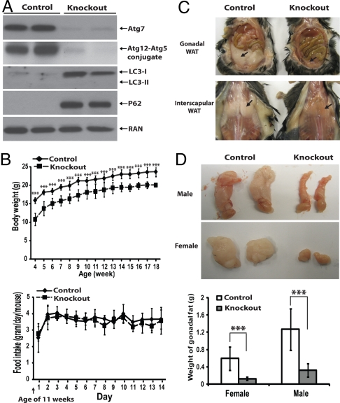
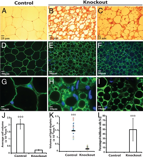
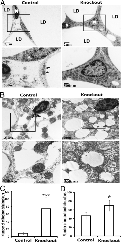
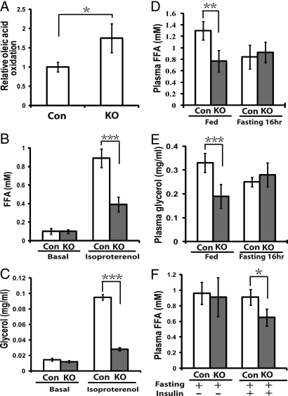
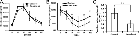
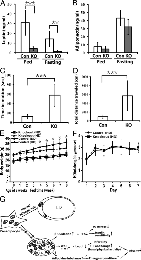
White adipocytes have a unique structure in which nearly the entire cell volume is occupied by one large lipid droplet. However, the molecular and cellular processes involved in the cytoplasmic remodeling necessary to create this structure are poorly defined. Autophagy is a membrane trafficking process leading to lysosomal degradation. Here, we investigated the effect of the deletion of an essential autophagy gene, autophagy-related gene 7 (atg7), on adipogenesis. A mouse model with a targeted deletion of atg7 in adipose tissue was generated. The mutant mice were slim and contained only 20% of the mass of white adipose tissue (WAT) found in wild-type mice. Interestingly, approximately 50% of the mutant white adipocytes were multilocular. The mutant white adipocytes were smaller with a larger volume of cytosol and contained more mitochondria. These cells exhibited altered fatty acid metabolism with increased rates of beta-oxidation and reduced rates of hormone-induced lipolysis. Consistently, the mutant mice had lower fed plasma concentrations of fatty acids and the levels decreased at faster rates upon insulin stimuli. These mutant mice exhibited increased insulin sensitivity. The mutant mice also exhibited markedly decreased plasma concentrations of leptin but not adiponectin, lower plasma concentrations of triglyceride and cholesterol, and they had higher levels of basal physical activity. Strikingly, these mutant mice were resistant to high-fat-diet-induced obesity. Taken together, our results indicate that atg7, and by inference autophagy, plays an important role in normal adipogenesis and that inhibition of autophagy by disrupting the atg7 gene has a unique anti-obesity and insulin sensitization effect.


Conflict of interest statement

The authors declare no conflict of interest.

Figures

Fig. 1.
Fig. 1.
Adipose- specific atg7 knockout mice exhibited reduced body weight and WAT mass. (A) Immunoblotting analyses of lysates of white adipose tissue (female, uterine WAT) from control (atg7 flox/flox) and adipose-specific atg7 conditional knockout (atg7flox/flox; ap2-Cre) mice using indicated antibodies (Atg12-Atg5 conjugate was detected with an anti-Atg12 antibody). (B) Upper, body weight chart for control (female, n = 12) and atg7 conditional knockout (female, n = 11) mice from 4–18 weeks. Lower, a 2-week food intake chart for control (female, n = 6) and atg7 conditional knockout (female, n = 6) mice starting from week 11. (C) Representative pictures of control and atg7 conditional knockout mice at the age of 20 weeks, showing gonadal (Upper) and interscapular (Lower) white adipose tissue (WAT) as indicated by arrows. (D) Representative pictures of gonadal fat pad (uterine fat in female and epididymal fat in male) and quantification (Lower) from control (male, n = 10; female, n = 12) and atg7 conditional knockout (male, n = 5; female, n = 6) mice at the age of 18–20 weeks. ***, P < 0.001, Student's t test.
Fig. 2.
Fig. 2.
Histological and immunofluorescence analysis of gonadal WAT from control and atg7 conditional knockout mice. (A–C) Representative microscopic pictures of H&E stained sections of uterine WAT from control (atg7flox/flox, A) and adipose-specific atg7 conditional knockout mice (atg7flox/flox; aP2-Cre, B and C). (D–I) Representative microscopic pictures of immunofluorescence assays of uterine WAT from control (D and G) and atg7 conditional knockout mice (E, F, H, and I) with Perilipin A antibody. D–F were pictures of low magnification and G–I were pictures of high magnification. (J–L) Quantification of average cell volume, lipid droplet volume, and percentage of multilocular cells, as indicated, of uterine WAT from control and atg7 conditional knockout mice. ***, P < 0.001, Student's t test. The data show representative results of tissues from six pairs of female mice (control and atg7 knockout).
Fig. 3.
Fig. 3.
Adipose-specific atg7 knockout mice accumulated more mitochondria in WAT and BAT (brown adipose tissue). (A) Electron microscopic pictures of adipocytes from uterine WAT of control and adipose-specific atg7 knockout mice. Selected regions in low magnification images (within the squares) are shown below in high magnification. LD, lipid droplet; N, nucleus; arrows indicate mitochondria in the control tissue sample. (B) Representative electron microscopic pictures of interscapular BAT from control and atg7 conditional knockout mice. Selected regions in low magnification images (within the squares) are shown below in high magnification. (C and D) Quantification of mitochondria in WAT and BAT of control and adipose-specific atg7 knockout mice, respectively. Mitochondria number was counted in 25 random cells and expressed as the number of mitochondria per nucleus. *, P < 0.05; ***, P < 0.001, Student's t test.
Fig. 4.
Fig. 4.
Fatty acid oxidation and lipolysis analysis of the adipose-specific atg7 conditional knockout mice. (A) β-oxidation analysis of white adipocytes isolated from control (CON, n = 3) and atg7 conditional knockout mice (KO, n = 4). 1-14C labeled oleic acid was added to the medium containing isolated adipocytes (5 × 10⁵), and 14CO2 trapped by the filter paper soaked with hyamine hydroxide was measured after a 3 h- incubation by a scintillation counter. (B and C) Lipolysis analysis of white adipocytes from control (CON, n = 3) and atg7 conditional knockout (KO, n = 4) mice. Adipocytes (5 × 10⁵) from control and atg7 conditional knockout mice were incubated in medium for 2 h in the absence or presence of 10 μM isoproterenol. Free fatty acid (FFA) (B) and glycerol (C) levels were then measured. (D) Plasma FFA levels of fed and fasting (16 h) mice (Con, n = 6; KO, n = 6). (E) Plasma glycerol levels of the fed and fasting (16 h) mice (Con, n = 6; KO, n = 6). (F) Plasma FFA levels of the mice (fasted for 5 h) before and 30 min after i.p. injection of insulin (0.75 U/kg, Con, n = 6; KO, n = 6). *, P < 0.05, **, P < 0.01, ***, P < 0.001. Student's t test. These data are representative results from two independent experiments.
Fig. 5.
Fig. 5.
Adipose- specific atg7 conditional knockout mice exhibited increased insulin sensitivity. (A) Glucose tolerance tests. Control mice (male, n = 6) and atg7 conditional knockout mice (male, n = 6) were fasted overnight before receiving an i.p. injection of 2 g/kg glucose and blood glucose concentration was measured at indicated time points. (B) Insulin tolerance tests. Control mice (male, n = 6) and atg7 conditional knockout (male, n = 6) mice were fasted for 5 h before receiving an i.p. injection of 0.75 U/kg insulin and blood glucose concentration were measured at indicated time points. (C) Plasma insulin levels of the control (male, n = 6) and atg7 conditional knockout mice (male, n = 6) fasted for 5 h. *, P < 0.05; **, P < 0.01, Student's t test. These data are representative results from two independent experiments.
Fig. 6.
Fig. 6.
The adipose-specific atg7 conditional knockout mice exhibited altered adipokine secretion, increased basal physical activity, and were resistant to high-fat diet induced obesity. (A) Leptin levels of the fed and fasted (5 h) control (n = 6) and atg7 conditional knockout mice (n = 6). (B) Adiponectin levels of the fed and fasted (5 h) control (n = 6) and atg7 conditional knockout mice (n = 6). The data from (A) and (B) are representative results from two independent experiments. (C and D) Open Field Test of mouse activity. 12 wild-type and 12 atg7 conditional knockout mice were analyzed with VersaMax animal activity monitor (Accuscan) for a period of 20 min. Time in motion (C) and total distance traveled (D) were recorded. (E) Body weight chart of control (atg7flox/flox, male, n = 9) and adipose- specific atg7 conditional knockout mice (atg7flox/flox;aP2-cre, male, n = 6) fed with normal diet (ND) or high fat diet (HD) from the age of 8–16 weeks. (F) One-week HD food intake chart of control (male, n = 6) and atg7 conditional knockout (male, n = 6) mice starting from week 14. (G) A working model summarizing the phenotypes of the adipose-specific atg7 knockout mice and the proposed underlying mechanisms. Detail was described in Discussion. *, P < 0.05, **, P < 0.01, ***, P < 0.001, Student's t test. LD, lipid droplet; N: nucleus; TG, triglyceride; ↑, upregulation; ↓, downregulation.

References

    1. Rosen ED, Spiegelman BM. Adipocytes as regulators of energy balance and glucose homeostasis. Nature. 2006;444:847–853. - PMC - PubMed
    1. Shulman GI. Cellular mechanisms of insulin resistance. J Clin Invest. 2000;106:171–176. - PMC - PubMed
    1. Friedman JM. Leptin at 14 y of age: An ongoing story. Am J Clin Nutr. 2009;89:973S–979S. - PMC - PubMed
    1. Farmer SR. Transcriptional control of adipocyte formation. Cell Metab. 2006;4:263–273. - PMC - PubMed
    1. Gesta S, Tseng YH, Kahn CR. Developmental origin of fat: Tracking obesity to its source. Cell. 2007;131:242–256. - PubMed

Publication types

MeSH terms