Comparative Study
. 2010 Jan;41(1):e34-40.
doi: 10.1161/STROKEAHA.109.568766. Epub 2009 Nov 12.

Reperfusion is a more accurate predictor of follow-up infarct volume than recanalization: a proof of concept using CT in acute ischemic stroke patients

Affiliations
Comparative Study

Reperfusion is a more accurate predictor of follow-up infarct volume than recanalization: a proof of concept using CT in acute ischemic stroke patients

Bruno P Soares et al. Stroke. 2010 Jan.

Abstract

Background and purpose: The purpose of this study was to compare recanalization and reperfusion in terms of their predictive value for imaging outcomes (follow-up infarct volume, infarct growth, salvaged penumbra) and clinical outcome in acute ischemic stroke patients.

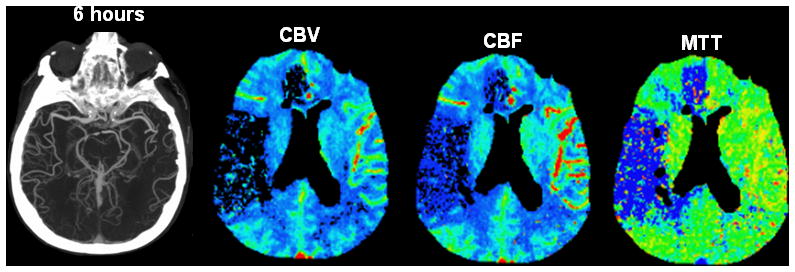
Material and methods: Twenty-two patients admitted within 6 hours of stroke onset were retrospectively included in this study. These patients underwent a first stroke CT protocol including CT-angiography (CTA) and perfusion-CT (PCT) on admission, and similar imaging after treatment, typically around 24 hours, to assess recanalization and reperfusion. Recanalization was assessed by comparing arterial patency on admission and posttreatment CTAs; reperfusion, by comparing the volumes of CBV, CBF, and MTT abnormality on admission and posttreatment PCTs. Collateral flow was graded on the admission CTA. Follow-up infarct volume was measured on the discharge noncontrast CT. The groups of patients with reperfusion, no reperfusion, recanalization, and no recanalization were compared in terms of imaging and clinical outcomes.

Results: Reperfusion (using an MTT reperfusion index >75%) was a more accurate predictor of follow-up infarct volume than recanalization. Collateral flow and recanalization were not accurate predictors of follow-up infarct volume. An interaction term was found between reperfusion and the volume of the admission penumbra >50 mL.

Conclusions: Our study provides evidence that reperfusion is a more accurate predictor of follow-up infarct volume in acute ischemic stroke patients than recanalization. We recommend an MTT reperfusion index >75% to assess therapy efficacy in future acute ischemic stroke trials that use perfusion-CT.


Figures

Fig. 1
Fig. 1. CBV, CBF and MTT reperfusion indices according to the recanalization status
Patients with recanalization had higher reperfusion indices than patients with no recanalization (CBV p = 0.16; CBF p = 0.04; MTT p = 0.18). The three black dots represent patients in which, although recanalization has been achieved, CBV and CBF reperfusion indices were very low or negative.
Fig. 2
Fig. 2. Absence of reperfusion, even in the setting of complete recanalization, may result in a large follow-up infarct volume
A. Axial noncontrast CT performed upon admission (2.5 hours after onset of symptoms) shows subtle hypoattenuation of the right putamen but no sulcal effacement in the right MCA territory. B. Axial maximum-intensity projection image from CTA performed upon admission shows occlusion of the M1 segment of the right MCA (arrow). PCT maps show decreased CBV and CBF in the right frontal and temporal lobes and a larger region of prolonged MTT that also involves the right ACA territory. The region of decreased CBV corresponds to the infarct core, whereas the surrounding mismatch region of prolonged MTT represents the ischemic penumbra. The patient received endovascular thrombectomy with a MERCI device. C. Axial maximum-intensity projection image from CTA and PCT performed 6 hours after admission show that, despite complete recanalization of the right MCA (arrow), PCT maps show that the region of decreased CBV and CBF has expanded to include the right anterior ACA territory that was previously considered tissue at risk. MTT is still abnormally increased in the right superficial MCA territory and in a portion of the right ACA territory. D. Axial noncontrast CT performed 48 hours after admission shows marked hypoattenuation and edema in the territories matching the perfusion deficit on the reperfusion PCT.
Fig. 3
Fig. 3. MTT reperfusion index according to recanalization status and collateral scores
Patients with recanalization had high MTT reperfusion indices, regardless of their collateral score. However, in patients with no recanalization, those with good collateral flow (score of 2 or 3) had a higher MTT reperfusion index than those with poor collateral flow (score of 0 or 1).
Fig. 4
Fig. 4. Imaging endpoints (follow-up infarct volume, infarct growth and salvaged penumbra) according to reperfusion and recanalization status
A. Follow-up infarct volume according to reperfusion and recanalization status. Patients with reperfusion had a smaller follow-up infarct volume than patients with no reperfusion, regardless of their recanalization status. B. Infarct growth according to reperfusion and recanalization status. Patients with reperfusion had smaller infarct growth than patients with no reperfusion, regardless of their recanalization status. C. Salvaged penumbra according to reperfusion and recanalization status. Patients with reperfusion had larger salvaged penumbra than patients with no reperfusion, regardless of their recanalization status.

Similar articles

Cited by

References

    1. Tomsick T. TIMI, TIBI, TICI: I came, I saw, I got confused. AJNR Am J Neuroradiol. 2007;28:382–384. - PMC - PubMed
    1. Tissue plasminogen activator for acute ischemic stroke. The National Institute of Neurological Disorders and Stroke rt-PA Stroke Study Group. N Engl J Med. 1995;333:1581–1587. - PubMed
    1. Zaidat OO, Suarez JI, Sunshine JL, Tarr RW, Alexander MJ, Smith TP, Enterline DS, Selman WR, Landis DM. Thrombolytic therapy of acute ischemic stroke: correlation of angiographic recanalization with clinical outcome. AJNR Am J Neuroradiol. 2005;26:880–884. - PMC - PubMed
    1. del Zoppo GJ, Koziol JA. Recanalization and stroke outcome. Circulation. 2007;115:2602–2605. - PubMed
    1. Rha JH, Saver JL. The impact of recanalization on ischemic stroke outcome: a meta-analysis. Stroke. 2007;38:967–973. - PubMed

Publication types